Alternate header for print version


Licensing
Public Domain: This image is in the public domain and thus free of any copyright restrictions. However, as is the norm in scientific publishing and as a matter of courtesy, any user should credit the content provider for any public or private use of this image whenever possible. Learn more
tweet  
*CIL – Cell Image Library accession number. Please use this to reference an image.

CIL:40165*  Cite 
Description

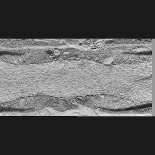
Single slice through a single tilt tomogram of the Node of Ranvier from mouse sciatic nerve prepared by high pressure freezing and freeze substitution of aldehyde fixed material. This image has been downsampled from the raw data image which can be accessed using the link provided to the Cell Centered Database. For more information see: Sosinsky et al. (2005) Development of a model for microphysiological simulations: small nodes of ranvier from peripheral nerves of mice reconstructed by electron tomography. Neuroinformatics. 3(2):133-62. PMID: 15988042

Technical Details

Tissue was obtained from a postnatal day 3 male mouse. Images were obtained using a JEOL 4000 microscope. Single tilt tomogram spanned -60 to 60° at 2° increments. Magnification: 25000X, accelerating voltage: 400.0 KeV.

Biological Sources
NCBI Organism Classification
Mus musculus
Cell Type
peripheral neuron
myelinating Schwann cell
Cellular Component
node of Ranvier
myelin sheath
Biological Context
Biological Process
regulation of action potential in neuron
Attribution
Names
Mark Ellisman
Gina Sosinsky
Link
CCDB 49
Citation
Digital Object Identifier (DOI)
doi:10.7295/W9CIL40165
Archival Resource Key (ARK)
ark:/b7295/w9cil40165
Grouping This image is part of a group.
Imaging
Image Type
recorded image
Image Mode
Parameters Imaged
electron density
Data Qualifiers
processed data
Sample Preparation
Methods
crosslinking-fixative fixed tissue
FBbi:00000621
Relation To Intact Cell
microtome-sectioned tissue
Dimensions
Spatial Axis Image Size Pixel Size
X 950px ——
Y 950px ——