Alternate header for print version
Advanced search
Contributors
Help
Submit
Search
menu
Cell Process
Cell Component
Cell Type
Organism
Microbial
Alzheimer's
Data Sets
University of California, San Diego
9500 Gilman Drive
La Jolla, CA 92093-0608, USA
Voice
: (858) 534-0276
Fax
: (858) 534-7497
Email
: dorloff@ncmir.ucsd.edu
Search Results for
neuron projection
(712 results)
(Not the results you were expecting?)
(Comments)
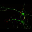
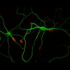
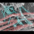
CIL:8787
NCBI Organism Classification
Rattus
Biological Process
developmental process
Cellular Component
cytoskeleton
This color combined image shows the spatial relationship between filamentous actin (red) and microtubule array (green) in cultured hippocampal neurons, grown for 1 day in vitro. Actin staining (with ...
CIL:8773
NCBI Organism Classification
Rattus
Biological Process
developmental process
Cellular Component
cytoskeleton
This color combined image shows the spatial relationship between filamentous actin (red) and microtubule array (green) in cultured hippocampal neurons, grown for 1 day in vitro. Actin staining (with ...
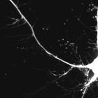
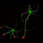
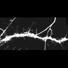
CIL:40810
NCBI Organism Classification
Rattus
Biological Process
dendrite morphogenesis
Cellular Component
axon
Colorized transmission electron micrograph of a platinum replica showing the cytoskeletal organization of stubby dendritic spines in extracted hippocampal neurons after 14 DIV. This imge shows branche...
CIL:37157
NCBI Organism Classification
Rattus
Biological Process
dendritic spine development
Cellular Component
dendritic spine
This is Video S2 which corresponds to still images in top row of Figure 2D. It shows the spine dynamics of control neurons. Time-lapse confocal imaging was performed on DIV14 hippocampal neurons co-ex...
CIL:37162
NCBI Organism Classification
Rattus
Biological Process
dendritic spine development
Cellular Component
dendritic spine
This is Video S3,which corresponds to still images in bottom row of Fig. 2D. It shows that knocking down levels of Myosin II B (MIIB) increases spine dynamics. Spines from MIIB knockdown neurons exten...
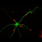
CIL:10221
NCBI Organism Classification
Rattus
Biological Process
developmental process
Cellular Component
cytoskeleton
This multi-layer image shows the spatial relationship between filamentous actin (red) and microtubule array (green) in cultured hippocampal neurons, grown for 3 days in vitro. Actin staining (with rh...
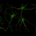
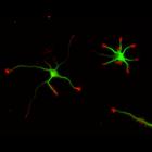
CIL:10226
NCBI Organism Classification
Rattus
Biological Process
developmental process
Cellular Component
cytoskeleton
This multi-layer image shows the spatial relationship between filamentous actin (red) and microtubule array (green) in cultured hippocampal neurons, grown for 3 days in vitro. Actin staining (with rh...
CIL:10230
NCBI Organism Classification
Rattus
Biological Process
developmental process
Cellular Component
cytoskeleton
This multi-layer image shows the spatial relationship between filamentous actin (red) and microtubule array (green) in cultured hippocampal neurons, grown for 5 days in vitro. Actin staining (with rh...
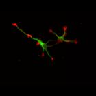
CIL:10235
NCBI Organism Classification
Rattus
Biological Process
developmental process
Cellular Component
cytoskeleton
This multi-layer image shows the spatial relationship between filamentous actin (red) and microtubule array (green) in cultured hippocampal neurons, grown for 5 days in vitro. Actin staining (with rh...
CIL:10236
NCBI Organism Classification
Rattus
Biological Process
developmental process
Cellular Component
cytoskeleton
This multi-layer image shows the spatial relationship between filamentous actin (red) and microtubule array (green) in cultured hippocampal neurons, grown for 5 days in vitro. Actin staining (with rh...
« Previous
1
...
3
4
5
6
7
8
9
10
...
72
Next »
Results per page:
10
20
50
100

")