Alternate header for print version
Advanced search
Contributors
Help
Submit
Search
menu
Cell Process
Cell Component
Cell Type
Organism
Microbial
Alzheimer's
Data Sets
University of California, San Diego
9500 Gilman Drive
La Jolla, CA 92093-0608, USA
Voice
: (858) 534-0276
Fax
: (858) 534-7497
Email
: dorloff@ncmir.ucsd.edu
Search Results for
Outer nuclear layer of retina
(14776 results)
(Not the results you were expecting?)
(Comments)
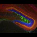
CIL:16245
NCBI Organism Classification
Mus musculus
Biological Process
cell communication by chemical coupling
Cellular Component
neuronal cell body membrane
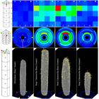
Distribution of interneurons expressing EGFP from the GAD65 promoter in the day 14 mouse hippocampus colabelled for the CB1 cannabinoid receptor (red) and counterstained with DAPI (blue) to show the c...
CCDB:8752
Species
mouse
Organ
eye
Cell type
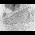
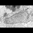
retina rod cell
System
central nervous system
Structure
mitochondrion
Cone and rod mitochondria: electron tomography
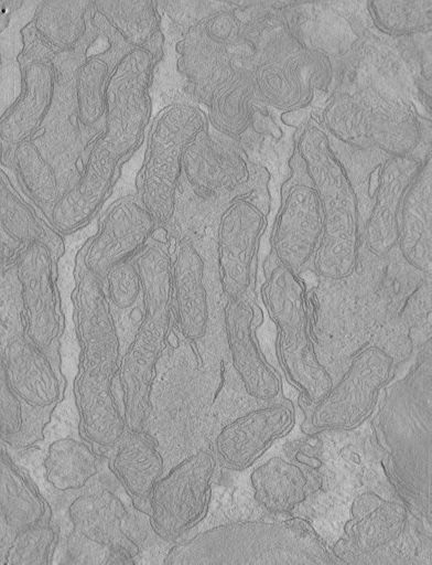
CIL:11458
NCBI Organism Classification
Homo sapiens
Biological Process
cellular respiration
Cellular Component
mitochondrion
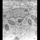
The inner segment of photoreceptors in the retina is packed with long, thin mitochondria that contain curved cristae. This image by Toichiro Kuwabara is of a photoreceptor from human retina, and appe...
CIL:50715
NCBI Organism Classification
Mus musculus
Biological Process
Native auditory Hair Cells at postnatal day 4
Cellular Component
Hair Cell stereocilia
FIB-SEM Dataset of anti-PKHD1L1 Immuno-Gold Labeled Outer Hair Cell Stereocilia Bundles. Postnatal day 4 mouse Inner Ear Organ of Corti labeled with anti-PKHD1L1 antibody (10 nm gold beads). The datas...
CIL:50728
NCBI Organism Classification
Mus musculus
Biological Process
Native auditory Hair Cells at postnatal day 4
Cellular Component
Hair Cell stereocilia
FIB-SEM Dataset of anti-PKHD1L1 Immuno-Gold Labeled Outer Hair Cell Stereocilia Bundles. Postnatal day 4 mouse Inner Ear Organ of Corti labeled with anti-PKHD1L1 antibody (10 nm gold beads). The datas...
CIL:38865
NCBI Organism Classification
Didinium nasutum
Biological Process
cell division
Cellular Component
cell division site
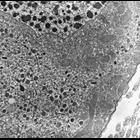
The division furrow of a dividing Didinium. A thick layer of epiplasm coated on the cytosolic side by mitochondria forms the constricting ring in the furrow. As usual mucocysts lie between the epiplas...
CIL:36665
NCBI Organism Classification
Paramecium multimicronucleatum
Biological Process
detection of symbiont
Cellular Component
symbiont-containing vacuole membrane
The first culture of P. multimicronucleatum which I had obtained from Carolina Biological Supply House contained a bacterial endosymbiont that resided specifically in the space between the two membran...
CIL:24922
NCBI Organism Classification
uncultured Scuticociliatia
Biological Process
cortical cytoskeleton organization
Cellular Component
macronucleus
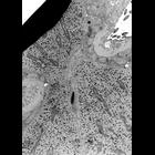
Ancistrum is a marine ciliate that inhabits the mantle cavity of mytiliid mussels. This image shows a good view of the macronucleus, mitochondria with tubular cristae, and what are probably flask-shap...
CIL:12090
NCBI Organism Classification
Paramecium tetraurelia
Biological Process
mitotic spindle elongation
Cellular Component
micronucleus
During division the micronucleus contains internal microtubules as well as a layer of b-tubulin, not in microtubular form, on or just inside the nuclear envelope. The section is immunogold labeled wit...
CIL:1314
NCBI Organism Classification
Paramecium tetraurelia
Biological Process
nuclear division
Cellular Component
micronucleus
High resolution view during division showing the micronucleus contains internal microtubules as well as a layer of b-tubulin, not in microtubular form, on or just inside the nuclear envelope. The sect...
« Previous
1
...
5
6
7
8
9
10
11
12
...
1478
Next »
Results per page:
10
20
50
100


")