Licensing
Description
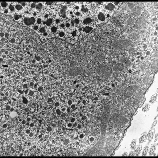
Ancistrum is a marine ciliate that inhabits the mantle cavity of mytiliid mussels. This image shows a good view of the macronucleus, mitochondria with tubular cristae, and what are probably flask-shaped endosomes. This group of images is located in a part of the cytoplasm that especially exhibits these vesicles, and CIL:24921 has a view at an appropriate angle to exhibit what may be dynamin or a dynamin-like protein decorating the tube of the flask shaped organelle. In addition, the macronuclear membrane is well demonstrated in this particular micrograph. TEM taken by G. Antipa in 1969 on a Hitachi HU11A operating at 75kV. The raw film was scanned with an Epson Perfection V750 Pro. This image is best used for quantitative analysis. Standard glutaraldehyde fixation followed by osmium tetroxide, dehydrated in alcohol and embedded in an epoxy resin. Microtome sections prepared at approximately 60nm thickness.
Biological Sources
- NCBI Organism Classification
- uncultured Scuticociliatia
- Ancistrum sp.
- Cell Type
- cell by organism
- eukaryotic cell
- Eukaryotic Protist
- Ciliated Protist
- Cellular Component
- macronucleus
- mitochondrial crista
- cytoplasm
- endosome
- cell cortex
- nuclear membrane
- nuclear outer membrane-endoplasmic reticulum membrane network
Biological Context
Attribution
- Name
- Gregory Antipa (San Francisco State University)
- Link
- http://online.sfsu.edu/~antipa/
Grouping
This image is part of a group.Imaging
- Image Type
- transmission electron microscopy (TEM)
- illumination by electrons
- Imaging Mode
- detection of electrons
- film
- Parameters Imaged
- electron density
- Source of Contrast
- stain with broad specificity
- Visualization Methods
- stain with broad specificity
- osmium tetroxide
- uranyl salt
- lead salt
- Processing History
- recorded image
- film
- unprocessed raw data
- Data Qualifiers
- raw, unprocessed data
- suitable for spatial measurements
Sample Preparation
- Methods
- glutaraldehyde fixed tissue
- osmium tetroxide fixed tissue
- tissue in epoxy resin embedment
- microtome-sectioned tissue
- Relation To Intact Cell
- microtome-sectioned tissue
Dimensions
| Spatial Axis | Image Size | Pixel Size |
|---|---|---|
| X | 5663px | 1.72nm |
| Y | 5561px | 1.72nm |