Alternate header for print version
Advanced search
Contributors
Help
Submit
Search
menu
Cell Process
Cell Component
Cell Type
Organism
Microbial
Alzheimer's
Data Sets
University of California, San Diego
9500 Gilman Drive
La Jolla, CA 92093-0608, USA
Voice
: (858) 534-0276
Fax
: (858) 534-7497
Email
: dorloff@ncmir.ucsd.edu
Search Results for
cellular localization
(714 results)
(Not the results you were expecting?)
(Comments)
CIL:13458
NCBI Organism Classification
Saccharomyces cerevisiae S288c
Biological Process
retrograde vesicle-mediated transport, Golgi to ER
Cellular Component
Golgi apparatus
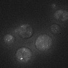
Intracellular localization of GFP-Vps74 in S. cerevisiae pik1-83 temperature-conditional strain grown at permissive temperature of 26C (control for 37C temperature-shift). In pik1-83 mutant after a 30...
CIL:13459
NCBI Organism Classification
Saccharomyces cerevisiae S288c
Biological Process
retrograde vesicle-mediated transport, Golgi to ER
Cellular Component
Golgi apparatus
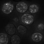
Intracellular localization of GFP-Vps74 in S. cerevisiae pik1-83 temperature-conditional strain grown at restrictive temperature of 37C. In pik1-83 mutant after a 30 minute incubation at the restricti...
CIL:24812
NCBI Organism Classification
Saccharomyces cerevisiae S288c
Biological Process
retrograde vesicle-mediated transport, Golgi to ER
Cellular Component
Golgi apparatus
Intracellular localization of GFP-Vps74 in BY4742 sac1∆ vps74∆. SAC1 encodes an integral membrane phosphoinositide phosphatase that is localized to the ER and Golgi. In the sac1∆ vps74∆ mutant...
CIL:24807
NCBI Organism Classification
Xenopus laevis
Biological Process
cellular localization
Cellular Component
lamellipodium
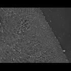
Localization of XAC (Xenopus ADF/cofilin) in Xenopus keratocytes done with immuno-EM. A higher mag view of a localized region of the cell is shown in CIL 24808. Image corresponds to Figure 8g from J...
CIL:24783
NCBI Organism Classification
Xenopus laevis
Biological Process
cellular localization
Cellular Component
lamellipodium
Localization of XAC (Xenopus ADF/cofilin) in Xenopus fibroblasts. Fluorescence microscopy of a cell fragment double stained with XAC antibody (green) and TRITC-phalloidin (red). XAC is distributed t...
CIL:24801
NCBI Organism Classification
Xenopus laevis
Biological Process
cellular localization
Cellular Component
lamellipodium
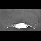
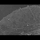
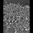
Structural differentiation of actin network in lamellipodium. Electron micrograph of Xenopus fibroblasts after regular extraction in the presence of polyethelene glycol (PEG) and phalloidin. While the...
CIL:24802
NCBI Organism Classification
Xenopus laevis
Biological Process
cellular localization
Cellular Component
lamellipodium
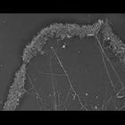
Structural differentiation of actin network in lamellipodium. Electon micrograph Xenopus fibroblast after unprotected extraction without polyethelene glycol. Actin network at lamellipodial rear disas...
CIL:24808
NCBI Organism Classification
Xenopus laevis
Biological Process
cellular localization
Cellular Component
lamellipodium
Localization of XAC (Xenopus ADF/cofilin) in Xenopus keratocytes done with immuno-EM. A low mag view of the cell from which this high mag view is taken is shown in CIL 24807. Image corresponds to Fi...
CIL:24809
NCBI Organism Classification
Xenopus laevis
Biological Process
cellular localization
Cellular Component
lamellipodium
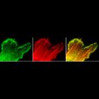
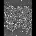
Localization of Xenopus ADF/cofilin (XAC) to posterior regions of depolymerization-resistant actin brush. Electron (micrograph of lamellipodia of Xenopus keratocytes after unprotected extraction and s...
CIL:24811
NCBI Organism Classification
Xenopus laevis
Biological Process
cellular localization
Cellular Component
lamellipodium
Localization of Xenopus ADF/cofilin (XAC) to posterior regions of depolymerization-resistant actin brush. Electron micrograph of lamellipodia of Xenopus keratocytes after latrunculin a treatment ( 0....
« Previous
1
...
6
7
8
9
10
11
12
13
...
72
Next »
Results per page:
10
20
50
100
")