Alternate header for print version
Advanced search
Contributors
Help
Submit
Search
menu
Cell Process
Cell Component
Cell Type
Organism
Microbial
Alzheimer's
Data Sets
Center for Research in Biological Systems
University of California, San Diego
9500 Gilman Drive
La Jolla, CA 92093-0608, USA
Voice
: (858) 534-0276
Fax
: (858) 534-7497
Email
: dorloff@ncmir.ucsd.edu
Search Results for
James D. Jamieson
(122 results)
(Not the results you were expecting?)
(Comments)
Still Images
Video/Animation
Z-Stack
Time Series
CIL:37131
NCBI Organism Classification
Rattus
Biological Process
regulation of vascular permeability
Cellular Component
vesicle
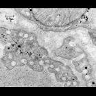
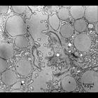
Transmission electron micrograph of rat capillary diaphragm 2h post intravenous administration of ferritin. Ferritin is seen entering plasmalemmal vesicles and into the extracellular space. Image mad...
CIL:37132
NCBI Organism Classification
Coloceras guinea
Biological Process
regulation of vascular permeability
Cellular Component
none specified
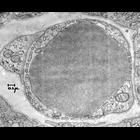
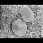
Transmission electron micrograph of a discontinuous endothelium in a capillary from a guinea pig pancreas. There is a red blood cell in the capillary lumen and a pericyte under the endothelium. Imag...
CIL:37138
NCBI Organism Classification
Cavia porcellus
Biological Process
exocytosis
Cellular Component
exocytic vesicle
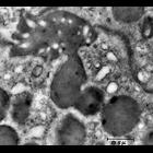
Transmission electron micrograph showing exocytosis in guinea pig pancreas. Image made available by James D. Jamieson and the Department of Cell Biology, Yale University School of Medicine.
CIL:37139
NCBI Organism Classification
Rattus
Biological Process
exocytosis
Cellular Component
exocytic vesicle
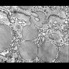
Transmission electron micrograph depicting exocytosis in a rat pancreas. Image made available by James D. Jamieson and the Department of Cell Biology, Yale University School of Medicine.
CIL:37140
NCBI Organism Classification
Rattus
Biological Process
exocytosis
Cellular Component
exocytic vesicle
Transmission electron micrograph showing a vesicle fusing with a duct lumen in a rat pancreas. Image made available by James D. Jamieson and the Department of Cell Biology, Yale University School of ...
CIL:37200
NCBI Organism Classification
Cavia porcellus
Biological Process
none specified
Cellular Component
mitochondrion
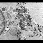
Transmission electron micrograph of guinea pig pancreas mitochondria and lipid droplets. The use of osmium tetroxide as a fixative for electron microscopy was first described by Dr. Palade at the Roc...
CIL:37202
NCBI Organism Classification
Cavia porcellus
Biological Process
none specified
Cellular Component
nucleus
Transmission electron micrograph of a section of the right atrium of a guinea pig heart. A portion of nucleus is seen at far left, with two centrioles close to the nuclear envelope. A region of myofib...
CIL:37207
NCBI Organism Classification
Rattus
Biological Process
regulation of cardiac muscle contraction
Cellular Component
intercalated disc
Transmission electron micrograph of longitudinal section of rat heart cardiac muscle from the atrium. A darkly stained intercalated disc runs from upper left to lower right. Also prominent are groups...
CIL:37209
NCBI Organism Classification
Rattus
Biological Process
regulation of cardiac muscle contraction
Cellular Component
atrial granuales
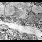
Transmission electron micrograph of longitudinal section of rat cardiac muscle from the heart atrium. Homogeneously staining spherical trial granules are present as well as mitochondria and myofibrils...
CIL:37212
NCBI Organism Classification
Rattus
Biological Process
regulation of cardiac muscle contraction
Cellular Component
mitochondrion
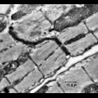
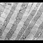
Transmission electron micrograph of longitudinal section of a a rat heart ventricle. Ordered arrays of myofibrils are interspersed with rows of mitochondria. Small, darkly staining glycogen granules a...
« Previous
1
...
6
7
8
9
10
11
12
13
Next »
Results per page:
10
20
50
100