Alternate header for print version
Advanced search
Contributors
Help
Submit
Search
menu
Cell Process
Cell Component
Cell Type
Organism
Microbial
Alzheimer's
Data Sets
Center for Research in Biological Systems
University of California, San Diego
9500 Gilman Drive
La Jolla, CA 92093-0608, USA
Voice
: (858) 534-0276
Fax
: (858) 534-7497
Email
: dorloff@ncmir.ucsd.edu
Search Results for
George E. Palade
(161 results)
(Not the results you were expecting?)
(Comments)
Still Images
Video/Animation
Z-Stack
Time Series
CIL:11180
NCBI Organism Classification
Rattus
Biological Process
maintenance of apical/basal cell polarity
Cellular Component
apical junction complex
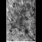
Electron micrograph of the junctional complex of intestinal epithelial cells of the rat shows the apical-most zonula occludens (tight junction), the zonula adherens (medium junction) and the macula ad...
CIL:7607
NCBI Organism Classification
Rattus rattus
Biological Process
biosynthetic process
Cellular Component
rough endoplasmic reticulum
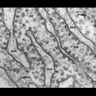
High magnification view of rough endoplasmic reticulum (RER) from rat pancreas showing RER and ribosomes, both bound (RiB) and free (RiF). Ribosomes were originally called Palade particles, as Palade ...
CIL:7587
NCBI Organism Classification
Rattus
Biological Process
lipid digestion
Cellular Component
lysosome
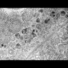
This image shows the heterogeneity in the content of lysosomes near the plasma membrane between two hepatocytes and near the bile canaliculus (not visible in this field). Some of the lysosomes contai...
CIL:37288
NCBI Organism Classification
Actinobacillus pleuropneumoniae
Biological Process
none specified
Cellular Component
none specified
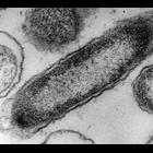
Transmission electron micrograph of bacteria hemophilus pleuropneumoniae. Image made available by James D. Jamieson and the Department of Cell Biology, Yale University School of Medicine.
CIL:37290
NCBI Organism Classification
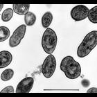
Streptococcus pneumoniae
Biological Process
none specified
Cellular Component
none specified
A transmission electron micrograph of a diplococcus (renamed Streptococcus in 1974) pneumoniae bacteria. Image made available by James D. Jamieson and the Department of Cell Biology, Yale University ...
CIL:37193
NCBI Organism Classification
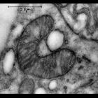
Plasmodium
Biological Process
none specified
Cellular Component
mitochondrion
Transmission electron micrograph of a mitochondria from the protist Plasmodium. The use of osmium tetroxide as a fixative for electron microscopy was first described by Dr. Palade at the Rockefeller....
CIL:37147
NCBI Organism Classification
none specified
Biological Process
none specified
Cellular Component
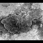
membrane
Transmission electron micrograph of cell fractionation artificial phopsholipid membranes. Image made available by James D. Jamieson and the Department of Cell Biology, Yale University School of Medic...
CIL:37195
NCBI Organism Classification
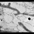
Neurospora
Biological Process
none specified
Cellular Component
mitochondrion
Transmission electron micrograph of a negatively stained mitochondria from the fungi Neurospora. The use of osmium tetroxide as a fixative for electron microscopy was first described by Dr. Palade at ...
CIL:37241
NCBI Organism Classification
none specified
Biological Process
none specified
Cellular Component
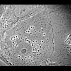
nucleus
This phase contrast image of a fibroblast was made available by James D. Jamieson and the Department of Cell Biology, Yale University School of Medicine.
CIL:37243
NCBI Organism Classification
none specified
Biological Process
none specified
Cellular Component
mitochondrion
Whole mount fibroblast: classic first images of endoplasmic reticulum and mitochondria. Image made available by James D. Jamieson and the Department of Cell Biology, Yale University School of Medicin...
« Previous
1
...
8
9
10
11
12
13
14
15
...
17
Next »
Results per page:
10
20
50
100