Alternate header for print version
Advanced search
Contributors
Help
Submit
Search
menu
Cell Process
Cell Component
Cell Type
Organism
Microbial
Alzheimer's
Data Sets
Center for Research in Biological Systems
University of California, San Diego
9500 Gilman Drive
La Jolla, CA 92093-0608, USA
Voice
: (858) 534-0276
Fax
: (858) 534-7497
Email
: dorloff@ncmir.ucsd.edu
Search Results for
Gary G. Borisy
(96 results)
(Not the results you were expecting?)
(Comments)
Still Images
Video/Animation
Z-Stack
Time Series
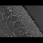
CIL:24800
NCBI Organism Classification
Xenopus laevis
Biological Process
actin filament organization
Cellular Component
lamellipodium
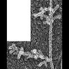
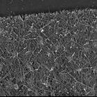
Structural differentiation of actin network in lamellipodium. Electron micrograph of Xenopus keratocyte after unprotected extraction without PEG and phalloidin. Actin network at lamellipodial rear di...
CIL:24803
NCBI Organism Classification
Xenopus laevis
Biological Process
cellular localization
Cellular Component
lamellipodium
Structural differentiation of actin network in lamellipodium. Electron micrograph of Xenopus fibroblast after unprotected extraction without polyethelene glycol and phalloidin. Actin network at lamel...
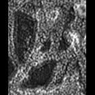
CIL:34891
NCBI Organism Classification
none specified
Biological Process
branching of actin filaments
Cellular Component
actin cytoskeleton
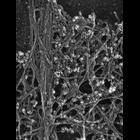
Multiple branching of actin filaments in lamellipodia of vertebrate fibroblasts. This image shows a local enlargement of the leading edge shown in overview in CIL 24788. Image corresponds to Figure 1...
CIL:34893
NCBI Organism Classification
none specified
Biological Process
branching of actin filaments
Cellular Component
actin cytoskeleton
Multiple branching of actin filaments in lamellipodia of vertebrate fibroblasts. This image shows a local enlargement of the leading edge shown in overview in CIL 24788. Image corresponds to Figure 1...
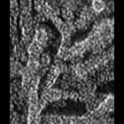
CIL:34901
NCBI Organism Classification
none specified
Biological Process
branching of actin filaments
Cellular Component
actin cytoskeleton
Improved visualization of actin filament branching in lamellipodia. EM of keratocyte or fibroblast lamellipodial actin network after cytochalasin D treatment (0.2 μM for 30 min or 0.5 μM for 10 min)...
CIL:24797
NCBI Organism Classification
none specified
Biological Process
actin filament-based process
Cellular Component
lamellipodium
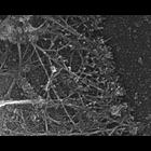
Localization of Arp2/3 complex in the lamellioidia of afibroblast . Staining with p21 antibody and 10 nm gold secondary antibody. CIL 24794 shows a fluorescence microsopy image of Arp2/3 localizatio...
CIL:24806
NCBI Organism Classification
Xenopus laevis
Biological Process
cellular localization
Cellular Component
lamellipodium
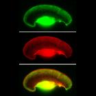
Localization of XAC (Xenopus ADF/cofilin) in Xenopus keratocytes. Fluorescence microscopy of a whole cell double stained with XAC antibody (green) and TRITC-phalloidin (red). XAC in lamellipodium is e...
CIL:24785
NCBI Organism Classification
Xenopus laevis
Biological Process
actin filament-based process
Cellular Component
lamellipodium
Localization of XAC (Xenopus ADF/cofilin) in Xenopus fibroblasts. Immuno-EM with XAC antibody at high magnification. Low magnification view is available at CIL 24784. Nucleus and surrounding regions...
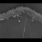
CIL:24786
NCBI Organism Classification
Xenopus laevis
Biological Process
branching of actin filaments
Cellular Component
lamellipodium
Multiple branching of actin filaments in lamellipodia of Xenopus keratocytes. This image shows an overview of the leading edge, and CIL 24787 shows enlargements of local regions of this platinum repli...
CIL:24792
NCBI Organism Classification
none specified
Biological Process
branching of actin filaments
Cellular Component
actin cytoskeleton
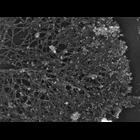
Electron micrograph of keratocyte or fibroblast lamellipodial actin network after latrunculin a (a chemical that binds monomeric actin) treatment (0.2 μM for 10 min). All examples demonstrate freque...
« Previous
1
2
3
4
5
6
7
8
9
10
Next »
Results per page:
10
20
50
100