Alternate header for print version
Advanced search
Contributors
Help
Submit
Search
menu
Cell Process
Cell Component
Cell Type
Organism
Microbial
Alzheimer's
Data Sets
Center for Research in Biological Systems
University of California, San Diego
9500 Gilman Drive
La Jolla, CA 92093-0608, USA
Voice
: (858) 534-0276
Fax
: (858) 534-7497
Email
: dorloff@ncmir.ucsd.edu
Search Results for
Don W. Fawcett
(332 results)
(Not the results you were expecting?)
(Comments)
Still Images
Video/Animation
Z-Stack
Time Series
CIL:35957
NCBI Organism Classification
Leporidae
Biological Process
fertilization
Cellular Component
flagellum
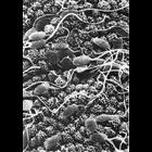
Figure 328 from Chapter 14 (Sperm Flagellum) of 'The Cell, 2nd Ed.' by Don W. Fawcett M.D. Scanning electron micrograph of rabbit spermatozoa on the endometrium of the uterus. Image by David Phillip...
CIL:10973
NCBI Organism Classification
Chinchilla
Biological Process
nucleus organization
Cellular Component
nucleus
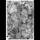
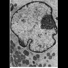
Transmission electron micrograph of a section of Chinchilla epididimus showing the highly lobed nucleus seen in Principal sells of the epididymal epithelium characteristic of many mammals. Figure 111...
CIL:10977
NCBI Organism Classification
Cavia porcellus
Biological Process
nucleus organization
Cellular Component
nuclear heterochromatin
Transmission electron micrograph showing the extensive accumulation of darkly stained heterochromatin at the nuclear periphery typical of many differentiated cells. Heterochromatin is excluded from re...
CIL:11027
NCBI Organism Classification
Didelphimorphia
Biological Process
nucleus organization
Cellular Component
nucleolus
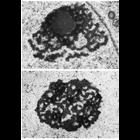
Transmission electron micrographs of Opossum spermatogonia showing different regions within the nucleolus. One region is more lightly staining and fine textured, the other denser and more coarsely tex...
CIL:11031
NCBI Organism Classification
Batrachoseps attenuatus
Biological Process
nucleus organization
Cellular Component
nucleolus
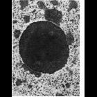
Transmission electron micrograph showing the highly compact nucleolus in the salamander hepatocyte. A finer textured central region is surrounded by a coarser region.
CIL:11037
NCBI Organism Classification
Sus scrofa scrofa
Biological Process
nucleus organization
Cellular Component
nucleus
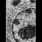
Transmission electron micrograph of Leydig cell nucleus from the domestic boar contains a region (arrow) thought to represent the single X chromosome which is largely heterochromatic. The chromosome i...
CIL:11046
NCBI Organism Classification
Cavia sp.
Biological Process
nucleus organization
Cellular Component
nuclear pore
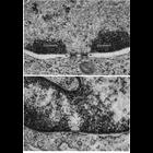
Transmission electron micrographs showing details of nuclear pores in thin sections of an erythroblast (above) and endothelial cell (below). Pores appear to be filled with dark staining material in th...
CIL:10987
NCBI Organism Classification
Unspecified
Biological Process
nucleus organization
Cellular Component
nucleus
Transmission electron micrograph of glutaraldehyde fixed pancreatic acinar cell showing the characteristic features of nuclear chromatin with this preparative method. Darkly staining heterochromatin (...
CIL:11014
NCBI Organism Classification
Ovis aries
Biological Process
nucleus organization
Cellular Component
nuclear chromosome
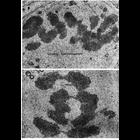
Transmission electron micrographs showing chromosomes in dividing spermatogonia during metaphase in tissue from ram testis (upper panel) and as they begin to separate during early anaphase in tissue f...
CIL:11023
NCBI Organism Classification
Homo sapiens
Biological Process
nucleus organization
Cellular Component
nuclear chromosome
Transmission electron micrograph showing part of a decondensed mitotic chromosome. The residual protein 'scaffold' (at bottom) is surrounded by a 'halo' of DNA loops 20 to 24 micrometers long. The en...
« Previous
1
2
3
4
5
6
7
8
9
...
34
Next »
Results per page:
10
20
50
100