Alternate header for print version
Advanced search
Contributors
Help
Submit
Search
menu
Cell Process
Cell Component
Cell Type
Organism
Microbial
Alzheimer's
Data Sets
Center for Research in Biological Systems
University of California, San Diego
9500 Gilman Drive
La Jolla, CA 92093-0608, USA
Voice
: (858) 534-0276
Fax
: (858) 534-7497
Email
: dorloff@ncmir.ucsd.edu
Search Results for
Don W Fawcett
(332 results)
(Not the results you were expecting?)
(Comments)
Still Images
Video/Animation
Z-Stack
Time Series
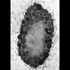
CIL:36008
NCBI Organism Classification
Homo sapiens
Biological Process
endocrine process
Cellular Component
secretory granule
Figure 390 from Chapter 15 (Cytoplasmic Inclusions) of 'The Cell, 2nd Ed.' by Don W. Fawcett M.D. Secretory granules of an alpha cell (upper part of the micrograph) and two beta cells (lower) from an ...
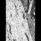
CIL:36009
NCBI Organism Classification
Rattus
Biological Process
secretory granule organization
Cellular Component
secretory granule
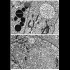
Figure 391 from Chapter 15 (Cytoplasmic Inclusions) of 'The Cell, 2nd Ed.' by Don W. Fawcett M.D. Secretory granules of a mast cell from rat connective tissue. Secretory granules in this cell type ar...
CIL:36014
NCBI Organism Classification
Rattus
Biological Process
synapse organization
Cellular Component
synapse
Figure 396 from Chapter 15 (Cytoplasmic Inclusions) of 'The Cell, 2nd Ed.' by Don W. Fawcett M.D. Axodendritic synapse from mammalian (rat) central nervous system. Arrow points to one region of the ...
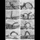
CIL:11135
NCBI Organism Classification
Felis catus
Biological Process
pinocytosis
Cellular Component
plasma membrane
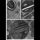
Electron micrographs show examples of pinocytosis in endothelial cells of blood vessels in the cat myocardium (panels A,B,C and H), and from the choroid rete of the bowfin fish eye. Both types of cell...
CIL:11150
NCBI Organism Classification
Homo sapiens
Biological Process
pinocytosis
Cellular Component
coated vesicle
This transmission electron micrograph of a section through a late orthochromatic erythroblast (normoblast) from human bone marrow shows shallow depressions along the membrane surface, indicated by arr...
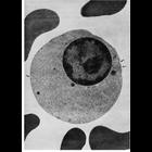
CIL:10837
NCBI Organism Classification
Perameles nasuta
Biological Process
autophagy
Cellular Component
lysosome
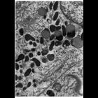
Figure 262 from Chapter 8 (Lysosomes) of 'The Cell, 2nd Ed.' by Don W. Fawcett M.D. Aggregations of primary lysosomes in the supranuclear region of an epithelial cell from the epididymis of the bandi...
CIL:10849
NCBI Organism Classification
Sus scrofa
Biological Process
autophagy
Cellular Component
lysosome
Figure 274 from Chapter 8 (Lysosomes) of 'The Cell, 2nd Ed.' by Don W. Fawcett M.D. Lysosomes in interstitial Leydig cells of the testis from the domestic boar, Sus scrofa. The laminar inclusions ap...
CIL:10850
NCBI Organism Classification
Chinchilla
Biological Process
phagocytosis
Cellular Component
lysosome
Figure 275 from Chapter 8 (Lysosomes) of 'The Cell, 2nd Ed.' by Don W. Fawcett M.D. Lysosomes of cells engaged in heterophagy, like the Sertoli cells from the testis of the chinchilla shown here, are...
CIL:10853
NCBI Organism Classification
Homo sapiens
Biological Process
endocytosis
Cellular Component
multivesicular body
Figure 277 from Chapter 8 (Lysosomes) of 'The Cell, 2nd Ed.' by Don W. Fawcett M.D. Multivesicular bodies, coated vesicles, and lysosomes are evident in these principle cells of the epithelial lining...
CIL:10855
NCBI Organism Classification
Homo sapiens
Biological Process
regulation of multivesicular body size
Cellular Component
multivesicular body
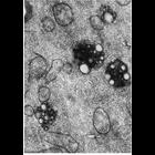
Figures 278 (upper) and 279 (lower) from Chapter 8 (Lysosomes) of 'The Cell, 2nd Ed.' by Don W. Fawcett M.D. Multivesicular bodies and lysososmes in human epididymal epithelium. Upper panel, arrows i...
« Previous
1
...
9
10
11
12
13
14
15
16
...
34
Next »
Results per page:
10
20
50
100