Alternate header for print version
Advanced search
Contributors
Help
Submit
Search
menu
Cell Process
Cell Component
Cell Type
Organism
Microbial
Alzheimer's
Data Sets
Center for Research in Biological Systems
University of California, San Diego
9500 Gilman Drive
La Jolla, CA 92093-0608, USA
Voice
: (858) 534-0276
Fax
: (858) 534-7497
Email
: dorloff@ncmir.ucsd.edu
Search Results for
David Sabatini
(30 results)
(Not the results you were expecting?)
(Comments)
Still Images
Video/Animation
Z-Stack
Time Series
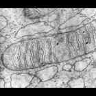
CIL:37199
NCBI Organism Classification
Cavia porcellus
Biological Process
none specified
Cellular Component
mitochondrion
Transmission electron micrograph of mitochondria from guinea pig pancreas. The use of osmium tetroxide as a fixative for electron microscopy was first described by Dr. Palade at the Rockefeller. Anot...
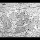
CIL:37196
NCBI Organism Classification
Cavia porcellus
Biological Process
none specified
Cellular Component
mitochondrion
Transmission electron micrograph of mitochondria from a guinea pig uterus. The use of osmium tetroxide as a fixative for electron microscopy was first described by Dr. Palade at the Rockefeller. Anot...
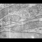
CIL:37194
NCBI Organism Classification
Cavia porcellus
Biological Process
none specified
Cellular Component
mitochondrion
Transmission electron micrograph of a mitochondria and a lipid droplet from a guinea pig pancreas. The use of osmium tetroxide as a fixative for electron microscopy was first described by Dr. Palade a...
CIL:37198
NCBI Organism Classification
Cavia porcellus
Biological Process
none specified
Cellular Component
mitochondrion
Transmission electron micrograph of mitochondria and mitochondrial granules from guinea pig pancreas. The use of osmium tetroxide as a fixative for electron microscopy was first described by Dr. Palad...
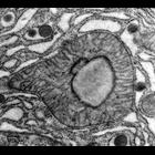
CIL:32137
NCBI Organism Classification
Drosophila melanogaster
Biological Process
cellular localization
Cellular Component
nucleus
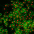
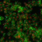
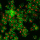
Drosophila melanogaster Kc167 cells were stained for DNA (to label nuclei, red) and actin (a cytoskeletal protein, to show the cell body, green). Each image is a dual channel fluorescent image followe...
CIL:32144
NCBI Organism Classification
Drosophila melanogaster
Biological Process
cellular localization
Cellular Component
nucleus
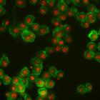
Drosophila melanogaster Kc167 cells were stained for DNA (to label nuclei, red) and actin (a cytoskeletal protein, to show the cell body, green). Each image is a dual channel fluorescent image followe...
CIL:32135
NCBI Organism Classification
Drosophila melanogaster
Biological Process
cellular localization
Cellular Component
nucleus
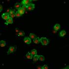
Drosophila melanogaster Kc167 cells were stained for DNA (to label nuclei, red) and actin (a cytoskeletal protein, to show the cell body, green). Each image is a dual channel fluorescent image followe...
CIL:32146
NCBI Organism Classification
Drosophila melanogaster
Biological Process
cellular localization
Cellular Component
nucleus
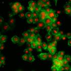
Drosophila melanogaster Kc167 cells were stained for DNA (to label nuclei, red) and actin (a cytoskeletal protein, to show the cell body, green). Each image is a dual channel fluorescent image followe...
CIL:32134
NCBI Organism Classification
Drosophila melanogaster
Biological Process
cellular localization
Cellular Component
nucleus
Drosophila melanogaster Kc167 cells were stained for DNA (to label nuclei, red) and actin (a cytoskeletal protein, to show the cell body, green). Each image is a dual channel fluorescent image followe...
CIL:32136
NCBI Organism Classification
Drosophila melanogaster
Biological Process
cellular localization
Cellular Component
nucleus
Drosophila melanogaster Kc167 cells were stained for DNA (to label nuclei, red) and actin (a cytoskeletal protein, to show the cell body, green). Each image is a dual channel fluorescent image followe...
« Previous
1
2
3
Next »
Results per page:
10
20
50
100