Alternate header for print version
Advanced search
Contributors
Help
Submit
Search
menu
Cell Process
Cell Component
Cell Type
Organism
Microbial
Alzheimer's
Data Sets
Center for Research in Biological Systems
University of California, San Diego
9500 Gilman Drive
La Jolla, CA 92093-0608, USA
Voice
: (858) 534-0276
Fax
: (858) 534-7497
Email
: dorloff@ncmir.ucsd.edu
Search Results for
Actin binding
(135 results)
(Not the results you were expecting?)
(Comments)
Still Images
Video/Animation
Z-Stack
Time Series
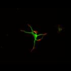
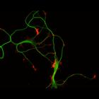
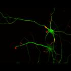
CIL:10209
NCBI Organism Classification
Rattus
Biological Process
developmental process
Cellular Component
cytoskeleton
This multi-layer image shows the spatial relationship between filamentous actin (red) and microtubule array (green) in cultured hippocampal neurons, grown for 1 day in vitro. Actin staining (with rho...
CIL:10213
NCBI Organism Classification
Rattus
Biological Process
developmental process
Cellular Component
cytoskeleton
This multi-layer image shows the spatial relationship between filamentous actin (red) and microtubule array (green) in cultured hippocampal neurons, grown for 1 day in vitro. Actin staining (with rho...
CIL:10215
NCBI Organism Classification
Rattus
Biological Process
developmental process
Cellular Component
cytoskeleton
This multi-layer image shows the spatial relationship between filamentous actin (red) and microtubule array (green) in cultured hippocampal neurons, grown for 1 day in vitro. Actin staining (with rho...
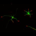
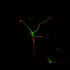
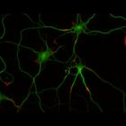
CIL:10220
NCBI Organism Classification
Rattus
Biological Process
developmental process
Cellular Component
cytoskeleton
This multi-layer image shows the spatial relationship between filamentous actin (red) and microtubule array (green) in cultured hippocampal neurons, grown for 3 days in vitro. Actin staining (with rh...
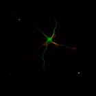
CIL:10228
NCBI Organism Classification
Rattus
Biological Process
developmental process
Cellular Component
cytoskeleton
This multi-layer image shows the spatial relationship between filamentous actin (red) and microtubule array (green) in cultured hippocampal neurons, grown for 5 days in vitro. Actin staining (with rh...
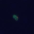
CIL:37908
NCBI Organism Classification
Homo sapiens
Biological Process
apoptotic process
Cellular Component
cortical actin cytoskeleton
Extrusion of an apoptotic cell from an HBE monolayer as shown by confocal Z-series. This set of images shows early extrusion events (middle and late events are CIL# 37909 and 37910). The bioactive lip...
CIL:8782
NCBI Organism Classification
Rattus
Biological Process
developmental process
Cellular Component
cytoskeleton
This color combined image shows the spatial relationship between filamentous actin (red) and microtubule array (green) in cultured hippocampal neurons, grown for 1 day in vitro. Actin staining (with ...
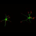
CIL:8775
NCBI Organism Classification
Rattus
Biological Process
developmental process
Cellular Component
cytoskeleton
This color combined image shows the spatial relationship between filamentous actin (red) and microtubule array (green) in cultured hippocampal neurons, grown for 1 day in vitro. Actin staining (with ...
CIL:10221
NCBI Organism Classification
Rattus
Biological Process
developmental process
Cellular Component
cytoskeleton
This multi-layer image shows the spatial relationship between filamentous actin (red) and microtubule array (green) in cultured hippocampal neurons, grown for 3 days in vitro. Actin staining (with rh...
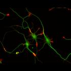
CIL:10226
NCBI Organism Classification
Rattus
Biological Process
developmental process
Cellular Component
cytoskeleton
This multi-layer image shows the spatial relationship between filamentous actin (red) and microtubule array (green) in cultured hippocampal neurons, grown for 3 days in vitro. Actin staining (with rh...
« Previous
1
...
7
8
9
10
11
12
13
14
Next »
Results per page:
10
20
50
100