Alternate header for print version
Advanced search
Contributors
Help
Submit
Search
menu
Cell Process
Cell Component
Cell Type
Organism
Microbial
Alzheimer's
Data Sets
University of California, San Diego
9500 Gilman Drive
La Jolla, CA 92093-0608, USA
Voice
: (858) 534-0276
Fax
: (858) 534-7497
Email
: dorloff@ncmir.ucsd.edu
Search Results for
visualization of contiguous regions
(2856 results)
(Not the results you were expecting?)
(Comments)
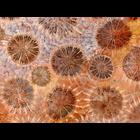
CIL:41842
NCBI Organism Classification
Corallium rubrum
Biological Process
none specified
Cellular Component
cell surface
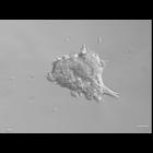
Fossil of red sponge coral, captured at 20x with brightfield microscopy. Honorable Mention, 2009 Olympus BioScapes Digital Imaging Competition®.
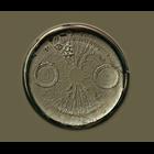
CIL:41826
NCBI Organism Classification
Bacillariophyta
Biological Process
unicellular algae organization
Cellular Component
frustule
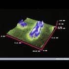
Differential interference contrast image of a diatom with tiny bits of shell from other diatoms on it. Honorable Mention, 2010 Olympus BioScapes Digital Imaging Competition®.
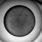
CIL:35275
NCBI Organism Classification
Danio rerio
Biological Process
embryo development
Cellular Component
cell
Zebrafish development from extension of head and tail rudiment during axiation, somitogenesis. Video made avaialable through Mark Cooper and Zebrafish - The Living Laboratory.
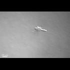
CIL:42153
NCBI Organism Classification
Mus musculus
Biological Process
dissemination
Cellular Component
cell surface
Representative time-lapse movie of a normal mouse mammary fragment in collagen I. CIL 42152 is related movie of a tumor mouse mammary fragment in collagen I. Images taken every 20 min. This movie i...
CIL:42156
NCBI Organism Classification
Mus musculus
Biological Process
dissemination
Cellular Component
cell surface
P-cadherin null epithelium shows enhanced and sustained dissemination of myoepithelial cells. Images taken every 20 minutes. This movie is part of a group of movies that include CIL 42151-42168.
CIL:39013
NCBI Organism Classification
Homo sapiens
Biological Process
mitotic metaphase
Cellular Component
chromosome
Fragile X chromosome made visible by atomic force microscopy (AFM). The arrow indicates the fragile site. Additional AFM chromosome images are available as CIL 38987, 39000, and 38915.
CIL:39089
NCBI Organism Classification
Plasmodium yoelii nigeriensis
Biological Process
sporozoan zygote development
Cellular Component
cyst wall
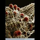
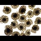
Colorized scanning electron micrograph of malaria (Plasmodium yoelii nigeriensis) oocysts ( thick-walled structure in which sporozoan zygotes develop) developing on the midgut wall of the mosquito Ano...
CIL:39107
NCBI Organism Classification
none specified
Biological Process
cell-cell adhesion
Cellular Component
cell surface
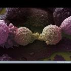
Lung cancer cells.
CIL:38915
NCBI Organism Classification
Homo sapiens
Biological Process
mitotic metaphase
Cellular Component
chromosome
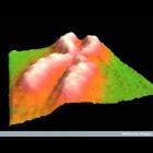
Scanning probe/atomic force microscope image of human chromosome 1. Additional scanning probe chromosome images are available as CIL 38987, 39000, and 39013.
CIL:41569
NCBI Organism Classification
Elphidium crispum
Biological Process
cell surface organization
Cellular Component
cell surface
Brightfield image of protozoan Elphidium crispum found growing on the Dorset coast of England. Honorable Mention, 2011 Olympus BioScapes Digital Imaging Competition®.
« Previous
1
...
13
14
15
16
17
18
19
20
...
286
Next »
Results per page:
10
20
50
100