Alternate header for print version
Advanced search
Contributors
Help
Submit
Search
menu
Cell Process
Cell Component
Cell Type
Organism
Microbial
Alzheimer's
Data Sets
University of California, San Diego
9500 Gilman Drive
La Jolla, CA 92093-0608, USA
Voice
: (858) 534-0276
Fax
: (858) 534-7497
Email
: dorloff@ncmir.ucsd.edu
Search Results for
whole mounted tissue
(2808 results)
(Not the results you were expecting?)
(Comments)
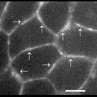
CIL:13884
NCBI Organism Classification
Saccharomyces cerevisiae
Biological Process
regulation of exit from mitosis
Cellular Component
nucleus
Constitutive targeting of Tem1 to the spindle pole body (SPB) in metaphase cells (this image) and anaphase cells (CIL#13887). tem1Δ::GAL-UPL-TEM1 cells expressing eGFP-BFA1–TEM1 from a CEN plasmid ...
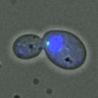
CIL:13890
NCBI Organism Classification
Saccharomyces cerevisiae
Biological Process
cell cycle
Cellular Component
nucleus
3HA-Bfa1 (red) is generally localized to one spindle pole body (SPB) in wild-type cells in metaphase as determined by spindle morphology (tubulin, green) and nuclear morphology (DAPI, blue). Control i...
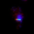
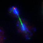
CIL:13892
NCBI Organism Classification
Saccharomyces cerevisiae
Biological Process
cell cycle
Cellular Component
nucleus
3HA-Bfa1 (red) is generally localized to the daughter spindle pole body (SPB) in wild-type cells in anaphase as determined by spindle morphology (tubulin, green) and nuclear morphology (DAPI, blue). C...
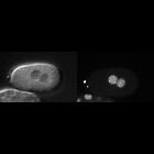
CIL:35149
NCBI Organism Classification
Caenorhabditis elegans
Biological Process
mitosis
Cellular Component
nucleus
This time-lapse series illustrates the early division up to the four-cell stage of a C. elegans embryo depleted of npl-4 by RNAi expressing H2B::GFP by DIC and fluorescence microscopy. GFP-tagged hist...
CIL:44509
NCBI Organism Classification
Canis lupus familiaris
Biological Process
wound healing, spreading of epidermal cells
Cellular Component
none specified
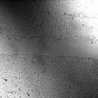
Time series light microscopy images illustrating a wound healing assay. A monolayer of MDCK (Madin-Darby Canine Kidney) epithelial cells is scratched to create a 'wound' about 300 micrometers in width...
CIL:43402
NCBI Organism Classification
Mus musculus
Biological Process
cell migration
Cellular Component
none specified
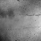
Wound Healing Assay Time series DIC images of mouse DA3 cells, derived from the mouse mammary adenocarcinoma cell line D1-DMBA-3, induced in BALB/C mice by dimethylbenzanthracene were grown to 90...
CIL:43403
NCBI Organism Classification
Mus musculus
Biological Process
cell migration
Cellular Component
none specified
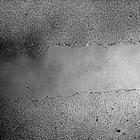
Wound Healing Assay Time series DIC images of mouse DA3 cells, derived from the mouse mammary adenocarcinoma cell line D1-DMBA-3, induced in BALB/C mice by dimethylbenzanthracene were grown to 90...
CIL:43405
NCBI Organism Classification
Mus musculus
Biological Process
cell migration
Cellular Component
none specified
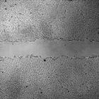
Wound Healing Assay Time series DIC images of mouse DA3 cells, derived from the mouse mammary adenocarcinoma cell line D1-DMBA-3, induced in BALB/C mice by dimethylbenzanthracene were grown to 90...
CIL:43417
NCBI Organism Classification
Mus musculus
Biological Process
cell migration
Cellular Component
none specified
Wound Healing Assay Time series DIC images of mouse DA3 cells, derived from the mouse mammary adenocarcinoma cell line D1-DMBA-3, induced in BALB/C mice by dimethylbenzanthracene were grown to 90...
CIL:24065
NCBI Organism Classification
Canis lupus familiaris
Biological Process
cell-cell adhesion
Cellular Component
adherens junction
E-cadherin local dynamics were studied in mature junctions, that is, junctions engaged in adhesion for many hours, in which cadherin expression level is stable. After stable transfection with E-cadher...
« Previous
1
...
273
274
275
276
277
278
279
280
281
Next »
Results per page:
10
20
50
100


")