Alternate header for print version
Advanced search
Contributors
Help
Submit
Search
menu
Cell Process
Cell Component
Cell Type
Organism
Microbial
Alzheimer's
Data Sets
University of California, San Diego
9500 Gilman Drive
La Jolla, CA 92093-0608, USA
Voice
: (858) 534-0276
Fax
: (858) 534-7497
Email
: dorloff@ncmir.ucsd.edu
Search Results for
optical path length gradient
(1067 results)
(Not the results you were expecting?)
(Comments)
CIL:34896
NCBI Organism Classification
none specified
Biological Process
branching of actin filaments
Cellular Component
actin cytoskeleton
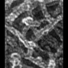
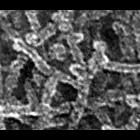
Improved visualization of actin filament branching in lamellipodia. EM of keratocyte or fibroblast lamellipodial actin network after cytochalasin D treatment (0.2 μM for 30 min or 0.5 μM for 10 min)...
CIL:34900
NCBI Organism Classification
none specified
Biological Process
branching of actin filaments
Cellular Component
actin cytoskeleton
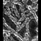
Improved visualization of actin filament branching in lamellipodia. EM of keratocyte or fibroblast lamellipodial actin network after cytochalasin D treatment (0.2 μM for 30 min or 0.5 μM for 10 min)...
CIL:34882
NCBI Organism Classification
Xenopus laevis
Biological Process
branching of actin filaments
Cellular Component
actin cytoskeleton
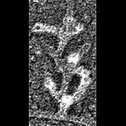
Multiple branching of actin filaments in lamellipodia of Xenopus keratocytes. This image shows an enlargement of a local region from the overview of the leading edge, CIL 24786. Image corresponds to ...
CIL:34884
NCBI Organism Classification
Xenopus laevis
Biological Process
branching of actin filaments
Cellular Component
actin cytoskeleton
Multiple branching of actin filaments in lamellipodia of Xenopus keratocytes. This image show an enlargement of a local region from the overview of the leading edge, CIL 24786. Image corresponds to F...
CIL:34886
NCBI Organism Classification
Xenopus laevis
Biological Process
branching of actin filaments
Cellular Component
actin cytoskeleton
Multiple branching of actin filaments in lamellipodia of Xenopus keratocytes. This image show an enlargement of a local region from the overview of the leading edge, CIL 24786. Image corresponds to F...
CIL:34888
NCBI Organism Classification
none specified
Biological Process
branching of actin filaments
Cellular Component
actin cytoskeleton
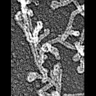
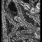
Multiple branching of actin filaments in lamellipodia of vertebrate fibroblasts. This image shows a local enlargement of the leading edge shown in overview in CIL 24788. Image corresponds to Figure 1...
CIL:37314
NCBI Organism Classification
Cavia porcellus
Biological Process
none specified
Cellular Component
cell surface
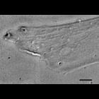
Nomaski image of live guinea pig pancreatic acinar cells. Image corresponds to Figure 1 in Proc Natl Acad Sci U S A. 1972 Oct;69(10):3028-32. Image made available by James D. Jamieson and the Departm...
CIL:38603
NCBI Organism Classification
Homo sapiens
Biological Process
none specified
Cellular Component
cell
This differential interference contrast image (DIC) corresponds to the same image field as the total internal reflection (TIRF) image CIL 38604 and the 2-color photoactivation localization microscopy ...
CIL:39466
NCBI Organism Classification
Ascidiacea
Biological Process
embryo development
Cellular Component
cell surface
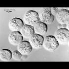
Early ascidian (sea squirt) embryos visualized by differential interface contrast (DIC) microscopy. Ascidians are used as a model for developmental research. Their simple embryonic development is rapi...
CIL:39471
NCBI Organism Classification
Ascidiacea
Biological Process
embryo development
Cellular Component
cell surface
Early ascidian (sea squirt) embryos visualized by differential interface contrast (DIC) microscopy. Ascidians are used as a model for developmental research. Their simple embryonic development is rapi...
« Previous
1
...
10
11
12
13
14
15
16
17
...
107
Next »
Results per page:
10
20
50
100