Alternate header for print version
Advanced search
Contributors
Help
Submit
Search
menu
Cell Process
Cell Component
Cell Type
Organism
Microbial
Alzheimer's
Data Sets
University of California, San Diego
9500 Gilman Drive
La Jolla, CA 92093-0608, USA
Voice
: (858) 534-0276
Fax
: (858) 534-7497
Email
: dorloff@ncmir.ucsd.edu
Search Results for
phase contrast microscopy
(951 results)
(Not the results you were expecting?)
(Comments)
CIL:35463
NCBI Organism Classification
Rattus norvegicus
Biological Process
none specified
Cellular Component
none specified
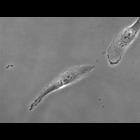
Phase contrast microscopy of liver sinusoidal endothelial cells purified from a Brown Rat.
CIL:35462
NCBI Organism Classification
Rattus norvegicus
Biological Process
none specified
Cellular Component
none specified
Phase contrast microscopy of purified liver sinusodial endothelial cells (LSECs) from Brown Rat.
CIL:54819
NCBI Organism Classification
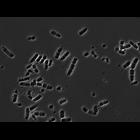
Alysiella filiformis
Biological Process
cell wall synthesis
Cellular Component
cell wall
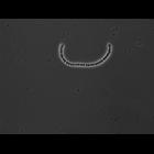
A. filiformis filament
CIL:54821
NCBI Organism Classification
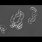
Alysiella filiformis
Biological Process
cell wall synthesis
Cellular Component
cell wall
A. filiformis filaments
CIL:54830
NCBI Organism Classification
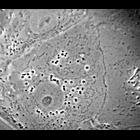
Alysiella filiformis
Biological Process
cell wall synthesis
Cellular Component
cell wall
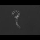
S. muelleri filaments
CIL:54834
NCBI Organism Classification
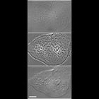
Simonsiella muelleri
Biological Process
cell wall synthesis
Cellular Component
cell wall
C. steedae filaments
CIL:37241
NCBI Organism Classification
none specified
Biological Process
none specified
Cellular Component
nucleus
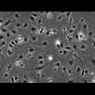
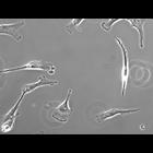
This phase contrast image of a fibroblast was made available by James D. Jamieson and the Department of Cell Biology, Yale University School of Medicine.
CIL:12594
NCBI Organism Classification
Homo sapiens
Biological Process
none specified
Cellular Component
cell
A human (Homo sapien) cheek cell smeared on a coverslip and imaged by birghtfield, phase contrast and differential interference contrast (DIC) microscopy. Images were acquired on a Zeiss Axiovert 200M...
CIL:25692
NCBI Organism Classification
Homo sapiens
Biological Process
mitosis
Cellular Component
none specified
Time lapse series of cell growth and division in cultured hTERT-RPE1 cells (telomerase immortalized human retinal pigment epithelium) using phase contrast optics. A movie created from this series is g...
CIL:25695
NCBI Organism Classification
Homo sapiens
Biological Process
mitosis
Cellular Component
none specified
Time lapse series of cell growth and division in cultured hTERT-RPE1 cells (telomerase immortalized human retinal pigment epithelium) using phase contrast optics. A movie created with this time series...
1
2
3
4
5
6
7
8
9
...
96
Next »
Results per page:
10
20
50
100


")