Alternate header for print version
Advanced search
Contributors
Help
Submit
Search
menu
Cell Process
Cell Component
Cell Type
Organism
Microbial
Alzheimer's
Data Sets
University of California, San Diego
9500 Gilman Drive
La Jolla, CA 92093-0608, USA
Voice
: (858) 534-0276
Fax
: (858) 534-7497
Email
: dorloff@ncmir.ucsd.edu
Search Results for
scanning electron microscopy (SEM)
(367 results)
(Not the results you were expecting?)
(Comments)
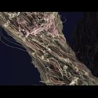
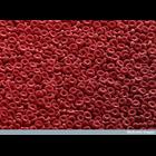
CIL:41738
NCBI Organism Classification
Homo sapiens
Biological Process
connective tissue organization
Cellular Component
fibrillar collagen
Colorized scanning electron micrograph of collagen/connective tissue removed from a human knee during arthroscopic surgery. The horizontal field width of the image is 16 microns. Wellcome Image Awar...
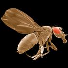
CIL:39726
NCBI Organism Classification
Drosophila melanogaster
Biological Process
sestrin null
Cellular Component
none specified
Scanning electron micrograph of a Drosophila melanogaster sestrin-null mutant. Sestrin-null Drosophila are used to study pathways involved in oxidative stress and aging. Relevant article: Lee, JH ...
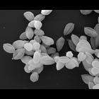
CIL:12403
NCBI Organism Classification
Leishmania mexicana
Biological Process
amastigote form Leishmania mexicana
Cellular Component
none specified
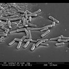
Leishmania mexicana amastigote forms (WHO strain MNYC/BZ/62/M379). Amastigotes were grown in axenic culture. The cells were fixed with glutaraldehyde, dehydrated in ethanol, critical-point dried and s...
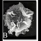
CIL:222
NCBI Organism Classification
none specified
Biological Process
none specified
Cellular Component
plasma membrane
A scanning electron microscope image of an activated mast cell illustrating the convoluted topography of the cell membrane, which is populated with receptors.
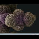
CIL:38811
NCBI Organism Classification
none specified
Biological Process
none specified
Cellular Component
cell surface
Scanning electron micrograph of red blood cells clearly showing their biconcave disc shape. Human red blood cells are typically 8 microns x 2 microns in size.
CIL:39104
NCBI Organism Classification
none specified
Biological Process
cell-cell adhesion
Cellular Component
cell surface
Scanning electron micrograph of pancreatic cancer cells. See additional image at CIL:39076.
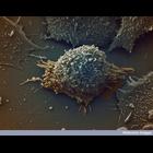
CIL:39106
NCBI Organism Classification
none specified
Biological Process
none specified
Cellular Component
cell surface
Colorized scanning electron micrograph of a lung cancer cell.
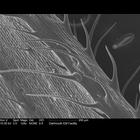
CIL:39358
NCBI Organism Classification
Zea mays
Biological Process
none specified
Cellular Component
none specified
Scanning electron microscope image of corn leaf surface.
CIL:38945
NCBI Organism Classification
none specified
Biological Process
carbohydrate biosynthetic process
Cellular Component
Golgi stack
Colorized scanning electron micrograph showing the stacked membrane discs of the Golgi complex. The Golgi is the area within a cell where many carbohydrates are synthesised, which can be used to modif...
CIL:40652
NCBI Organism Classification
none specified
Biological Process
response to bacterium
Cellular Component
cell surface
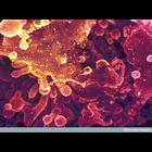
Scanning electron micrograph illustrating bacteria contamination of cells.
« Previous
1
...
3
4
5
6
7
8
9
10
...
37
Next »
Results per page:
10
20
50
100