Alternate header for print version
Advanced search
Contributors
Help
Submit
Search
menu
Cell Process
Cell Component
Cell Type
Organism
Microbial
Alzheimer's
Data Sets
University of California, San Diego
9500 Gilman Drive
La Jolla, CA 92093-0608, USA
Voice
: (858) 534-0276
Fax
: (858) 534-7497
Email
: dorloff@ncmir.ucsd.edu
Search Results for
scanning electron microscopy (SEM)
(367 results)
(Not the results you were expecting?)
(Comments)
CIL:39075
NCBI Organism Classification
none specified
Biological Process
none specified
Cellular Component
cell surface
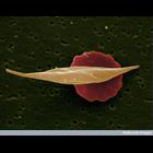
Scanning electron micrograph of normal red blood cell (background, colored red) and red blood cell affected by sickle-cell anaemia (foreground, colored tan). Sickle-cell anemia is a blood disease that...
CIL:39790
NCBI Organism Classification
Lytechinus pictus
Biological Process
embryonic morphogenesis
Cellular Component
cell surface
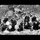
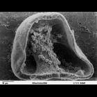
Scanning electron microscope image of Strongylocentrotus drobachiensus [sea urchin] gastrula. This is a higher magnification image of CIL39765. The embryo was split open to reveal a nice cross-section...
CIL:12663
NCBI Organism Classification
Danio rerio
Biological Process
none specified
Cellular Component
extracellular matrix
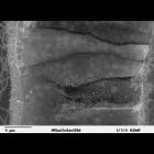
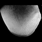
High magnification scanning EM of the extracellular matrix in the ventrolateral mesoderm of a 24 hr (prim-5) Zebrafish embryo. Specimens were chemically fixed critically point dried, and sputter coate...
CIL:39789
NCBI Organism Classification
Lytechinus pictus
Biological Process
syncytial ring formation
Cellular Component
cell surface
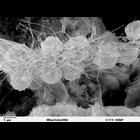
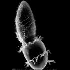
Scanning electron microscope image of Strongylocentrotus drobachiensus [sea urchin] embryo at the late gastrula stage. This is a high magnification image of CIL 39785. Embryo was cut to reveal blastoc...
CIL:39784
NCBI Organism Classification
Lytechinus pictus
Biological Process
cell migration
Cellular Component
cell surface
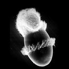
Scanning electron microscope image of Strongylocentrotus drobachiensus [sea urchin] at the gastrula stage. Embryo was cut in half to reveal blastocoel cavity, containing blastocoel matrix material, pr...
CIL:17891
NCBI Organism Classification
Didinium nasutum
Biological Process
phagocytosis
Cellular Component
oral apparatus
The ingestion of Paramecium by Didinium. This micrograph also clearly shows the five short rows of clavate cilia (ciliary stubs without central microtubules) that reside just below the anterior and po...
CIL:19535
NCBI Organism Classification
Didinium nasutum
Biological Process
phagocytosis
Cellular Component
oral apparatus
Didinium captures Paramecium. Also showing metachronous waves of cilia in the two characteristic ciliary girdles of Didinium nasutum. This micrograph was taken in 1968 by G. Antipa on a Cambridge Mar...
CIL:22783
NCBI Organism Classification
uncultured scuticociliate
Biological Process
cortical cytoskeleton organization
Cellular Component
cell cortex
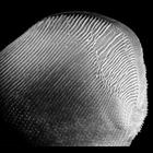
A view of the left side of Conchophthirus curtus deciliated by a calcium shock method to reveal the organization of the ciliature of this densely ciliated organism. In particular this method reveals t...
CIL:40905
NCBI Organism Classification
uncultured scuticociliate
Biological Process
cortical cytoskeleton organization
Cellular Component
cilium
Conchophthirus was deciliated by calcium ion shock followed by shearing through a micropipette. This revealed the location of the deciliated basal bodies and the cell's surface architecture. In this i...
CIL:40259
NCBI Organism Classification
Mus musculus
Biological Process
cerebellum structural organization
Cellular Component
synapse
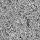
Single slice from a cropped and contrast-adjusted serial section reconstruction from the molecular layer of the cerebellar vermis of the adult mouse generated from serial block face SEM. This image h...
« Previous
1
...
25
26
27
28
29
30
31
32
...
37
Next »
Results per page:
10
20
50
100