Alternate header for print version
Advanced search
Contributors
Help
Submit
Search
menu
Cell Process
Cell Component
Cell Type
Organism
Microbial
Alzheimer's
Data Sets
University of California, San Diego
9500 Gilman Drive
La Jolla, CA 92093-0608, USA
Voice
: (858) 534-0276
Fax
: (858) 534-7497
Email
: dorloff@ncmir.ucsd.edu
Search Results for
scanning electron microscopy (SEM)
(367 results)
(Not the results you were expecting?)
(Comments)
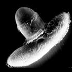
CIL:39247
NCBI Organism Classification
Didinium nasutum
Biological Process
phagocytosis
Cellular Component
oral apparatus
Didinium captures Paramecium. After reeling in the prey with cyclosis tugging at the attached toxicysts the proboscis opens to engage the Paramecium. This micrograph was taken in 1968 by G. Antipa on ...
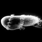
CIL:39249
NCBI Organism Classification
Didinium nasutum
Biological Process
phagocytosis
Cellular Component
oral apparatus
Didinium captures Paramecium. At midingestion and showing metachronous waves of cilia within the two characteristic ciliary girdles of Didinium nasutum. This micrograph was taken in 1968 by G. Antipa ...
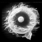
CIL:19536
NCBI Organism Classification
Didinium nasutum
Biological Process
oral apparatus organization
Cellular Component
oral apparatus
Apical oral view of Didinium. Showing the proboscis containing extrusive organelles to demobilize and eventually engulph it's prey. Metachronous waves of cilia in the anterior girdle of the two charac...
CIL:39782
NCBI Organism Classification
Lytechinus pictus
Biological Process
embryonic cleavage
Cellular Component
cleavage furrow
Scanning electron microscope image of Lytechinus pictus [sea urchin] embryo at the 2-cell stage. The fertilization envelope has been removed to reveal the cells covered with a dense meshwork of the hy...
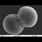
CIL:39791
NCBI Organism Classification
Lytechinus pictus
Biological Process
embryonic morphogenesis
Cellular Component
extracellular matrix
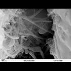
Scanning electron microscope image of Strongylocentrotus drobachiensus [sea urchin] embryo at the late gastrula stage. This high magnification image of the embryo shows the primary mesenchyme syncytia...
CIL:39787
NCBI Organism Classification
Lytechinus pictus
Biological Process
embryonic morphogenesis
Cellular Component
filopodium
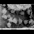
Scanning electron microscope image of Strongylocentrotus drobachiensus [sea urchin] at the gastrula stage. Embryo was split open to reveal the blastocoel cavity. There are several migrating mesenchyme...
CIL:39305
NCBI Organism Classification
Mus musculus
Biological Process
cell-cell junction organization
Cellular Component
cell-cell contact zone
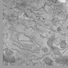
Focused ion beam scanning electron microscopy image series of a region of cell-cell contact between two cells in the multilayered region of an FGF2 (fibroblast growth factor 2) induced mammary organoi...
CIL:39057
NCBI Organism Classification
none specified
Biological Process
blood coagulation
Cellular Component
fibrin fibers
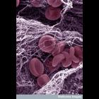
Colorized scanning electron micrograph of the components of a blood clot. Clotting of the blood is an important process that helps the body repair after injury. The blood is composed of red blood cell...
CIL:39783
NCBI Organism Classification
Lytechinus pictus
Biological Process
embryonic cleavage
Cellular Component
cell surface
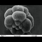
Scanning electron microscope image of Lytechinus pictus [sea urchin] embryo at the 16-cell stage. The four large macromeres are behind the four small micromeres. The eight mesomeres are behind the mac...
CIL:39788
NCBI Organism Classification
Lytechinus pictus
Biological Process
embryonic morphogenesis
Cellular Component
cell surface
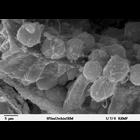
Scanning electron microscope image of Strongylocentrotus drobachiensus embryo at the primary mesenchyme blastula stage. Embryo was split open to reveal the outer epithelial layer and the blastocoel ca...
« Previous
1
...
24
25
26
27
28
29
30
31
...
37
Next »
Results per page:
10
20
50
100