Alternate header for print version
Advanced search
Contributors
Help
Submit
Search
menu
Cell Process
Cell Component
Cell Type
Organism
Microbial
Alzheimer's
Data Sets
University of California, San Diego
9500 Gilman Drive
La Jolla, CA 92093-0608, USA
Voice
: (858) 534-0276
Fax
: (858) 534-7497
Email
: dorloff@ncmir.ucsd.edu
Search Results for
neuron projection
(712 results)
(Not the results you were expecting?)
(Comments)
CIL:8785
NCBI Organism Classification
Rattus
Biological Process
developmental process
Cellular Component
cytoskeleton
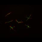
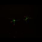
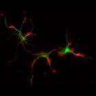
This color combined image shows the spatial relationship between filamentous actin (red) and microtubule array (green) in cultured hippocampal neurons, grown for 1 day in vitro. Actin staining (with ...
CIL:8780
NCBI Organism Classification
Rattus
Biological Process
developmental process
Cellular Component
cytoskeleton
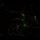
This color combined image shows the spatial relationship between filamentous actin (red) and microtubule array (green) in cultured hippocampal neurons, grown for 1 day in vitro. Actin staining (with ...
CIL:37154
NCBI Organism Classification
Rattus
Biological Process
dendritic spine development
Cellular Component
dendritic spine
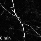
This is Video S1 and it corresponds to Figure 2A. It shows that local blebbistatin micropipetting, but not DMSO, increases spine formation and extension (arrowheads). Equal volumes of blebbistatin and...
CIL:38907
NCBI Organism Classification
Rattus
Biological Process
stem cell differentiation
Cellular Component
nucleus
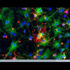
Confocal micrograph of astrocytes and oligodendrocytes from neural stem cells Astrocytes (green) and oligodendrocytes (red) derived from rat neural stem cells in culture. Both these cells are types of...
CIL:40804
NCBI Organism Classification
Rattus
Biological Process
dendrite morphogenesis
Cellular Component
axon
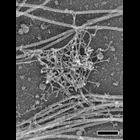
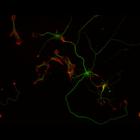
Colorized transmission electron micrograph of a platinum replica showing the cytoskeletal organization of stubby dendritic spines in extracted hippocampal neurons after 14 DIV. This image shows axons,...
CIL:36044
NCBI Organism Classification
Vertebrata
Biological Process
microtubule cytoskeleton organization
Cellular Component
microtubule
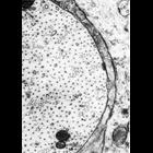
Figure 407 from Chapter 16 (Cytoplasmic matrix and cytoskeleton) of 'The Cell, 2nd Ed.' by Don W. Fawcett M.D. Proximal dendrite from a neuron of the anterior horn of the spinal cord. In this microg...
CIL:10095
NCBI Organism Classification
Rattus
Biological Process
developmental process
Cellular Component
cytoskeleton
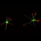
This multi-layer image shows the spatial relationship between filamentous actin (red) and microtubule array (green) in cultured hippocampal neurons, grown for 1 day in vitro. Actin staining (with rho...
CIL:10096
NCBI Organism Classification
Rattus
Biological Process
developmental process
Cellular Component
cytoskeleton
This multi-layer image shows the spatial relationship between filamentous actin (red) and microtubule array (green) in cultured hippocampal neurons, grown for 1 day in vitro. Actin staining (with rho...
CIL:10109
NCBI Organism Classification
Rattus
Biological Process
developmental process
Cellular Component
cytoskeleton
This multi-layer image shows the spatial relationship between filamentous actin (red) and microtubule array (green) in cultured hippocampal neurons, grown for 3 days in vitro. Actin staining (with rh...
CIL:10112
NCBI Organism Classification
Rattus
Biological Process
developmental process
Cellular Component
cytoskeleton
This multi-layer image shows the spatial relationship between filamentous actin (red) and microtubule array (green) in cultured hippocampal neurons, grown for 3 days in vitro. Actin staining (with rh...
« Previous
1
2
3
4
5
6
7
8
9
...
72
Next »
Results per page:
10
20
50
100

")