Alternate header for print version
Advanced search
Contributors
Help
Submit
Search
menu
Cell Process
Cell Component
Cell Type
Organism
Microbial
Alzheimer's
Data Sets
University of California, San Diego
9500 Gilman Drive
La Jolla, CA 92093-0608, USA
Voice
: (858) 534-0276
Fax
: (858) 534-7497
Email
: dorloff@ncmir.ucsd.edu
Search Results for
actin filament
(332 results)
(Not the results you were expecting?)
(Comments)
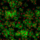
CIL:32132
NCBI Organism Classification
Drosophila melanogaster
Biological Process
cellular localization
Cellular Component
nucleus
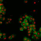
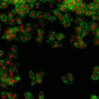
Drosophila melanogaster Kc167 cells were stained for DNA (to label nuclei, red) and actin (a cytoskeletal protein, to show the cell body, green). Each image is a dual channel fluorescent image followe...
CIL:32133
NCBI Organism Classification
Drosophila melanogaster
Biological Process
cellular localization
Cellular Component
nucleus
Drosophila melanogaster Kc167 cells were stained for DNA (to label nuclei, red) and actin (a cytoskeletal protein, to show the cell body, green). Each image is a dual channel fluorescent image followe...
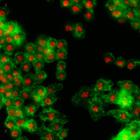
CIL:32139
NCBI Organism Classification
Drosophila melanogaster
Biological Process
cellular localization
Cellular Component
nucleus
Drosophila melanogaster Kc167 cells were stained for DNA (to label nuclei, red) and actin (a cytoskeletal protein, to show the cell body, green). Each image is a dual channel fluorescent image followe...
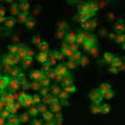
CIL:32140
NCBI Organism Classification
Drosophila melanogaster
Biological Process
cellular localization
Cellular Component
nucleus
Drosophila melanogaster Kc167 cells were stained for DNA (to label nuclei, red) and actin (a cytoskeletal protein, to show the cell body, green). Each image is a dual channel fluorescent image followe...
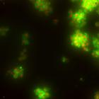
CIL:32141
NCBI Organism Classification
Drosophila melanogaster
Biological Process
cellular localization
Cellular Component
nucleus
Drosophila melanogaster Kc167 cells were stained for DNA (to label nuclei, red) and actin (a cytoskeletal protein, to show the cell body, green). Each image is a dual channel fluorescent image followe...
CIL:32142
NCBI Organism Classification
Drosophila melanogaster
Biological Process
cellular localization
Cellular Component
nucleus
Drosophila melanogaster Kc167 cells were stained for DNA (to label nuclei, red) and actin (a cytoskeletal protein, to show the cell body, green). Each image is a dual channel fluorescent image followe...
CIL:35563
NCBI Organism Classification
Rattus rattus
Biological Process
cell proliferation
Cellular Component
keratin filament
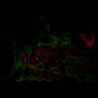
Liver of a rat exposed to chemical carcinogens. Serial sections of the liver imaged by laser scanning confocal microscopy. F-actin (red) is in bile canaliculi at junctions of the hepatocytes, which ...
CIL:36278
NCBI Organism Classification
Vorticella convallaria
Biological Process
contractile vacuole organization
Cellular Component
contractile vacuole pore
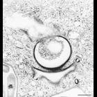
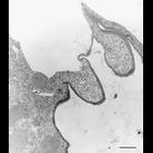
Contractile vacuole pore in face view illustrating the microtubular ribbons that extend from the pore to the CV membrane, the coil of microtubules that surrounds the pore and smaller pellicular pores ...
CIL:39339
NCBI Organism Classification
Nassula
Biological Process
contractile vacuole pore organization
Cellular Component
contractile vacuole pore
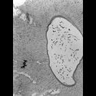
Detail of CV pore. Microtubules encircle the pore wall and other microtubules radiate out from the pore toward the CV membrane. TEM taken on 3/27/69 by R. Allen with Philips 300 operating at 60kV. Neg...
CIL:9907
NCBI Organism Classification
Euplotes sp.
Biological Process
water transport
Cellular Component
contractile vacuole pore
A rare micrograph of the contractile vacuole and intact contractile vacuole pore membrane of Euplotes. Close examination at high magnification reveals the organization of microtubules lining the pore ...
« Previous
1
...
26
27
28
29
30
31
32
33
34
Next »
Results per page:
10
20
50
100
")