Alternate header for print version
Advanced search
Contributors
Help
Submit
Search
menu
Cell Process
Cell Component
Cell Type
Organism
Microbial
Alzheimer's
Data Sets
University of California, San Diego
9500 Gilman Drive
La Jolla, CA 92093-0608, USA
Voice
: (858) 534-0276
Fax
: (858) 534-7497
Email
: dorloff@ncmir.ucsd.edu
Search Results for
mitotic metaphase
(121 results)
(Not the results you were expecting?)
(Comments)
CIL:15793
NCBI Organism Classification
Strongylocentrotus purpuratus
Biological Process
trichostatin A treatment
Cellular Component
microtubule
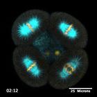
16-cell purple urchin embryo with EMTB-3G (microtubules) in shown cyan and mC-H2B (chromatin) in yellow. The embryo is treated with 15 µM trichostatin A (TSA), an inhibitor of HDAC6 (a tubulin deace...
CIL:40376
NCBI Organism Classification
Sus scrofa domestica
Biological Process
mitosis
Cellular Component
microtubule cytoskeleton
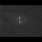
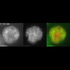
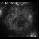
Time lapse movie of a porcine kidney LLCPK1 cell stably expressing photoactivable GFP-tubulin. Fluorescence was activated at metaphase in a vertical strip between the metaphase plate and spindle pole....
CIL:11992
NCBI Organism Classification
Drosophila melanogaster
Biological Process
mitotic anaphase
Cellular Component
myosin regulatory light chain
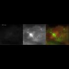
TIRF time lapse of mCherry-tubulin (left), myosin-GFP (middle), and overlay (myosin in green and tubulin in red; right) in a cell with a monopolar spindle from metaphase to anaphase. The S2 cells are...
CIL:35481
NCBI Organism Classification
Homo sapiens
Biological Process
mitotic metaphase
Cellular Component
spindle microtubule
Tripolar mitosis in liver cancer cell line. Normally, dividing cells form a single metaphase plate of paired chromosomes which are pulled apart by opposing microtubule spindles. This sequence shows p...
CIL:11987
NCBI Organism Classification
Drosophila melanogaster
Biological Process
mitotic anaphase
Cellular Component
myosin regulatory light chain
TIRF time lapse of myosin-GFP (left), mCherry-tubulin (middle), and overlay (myosin-GFP in green and mCherry-tubulin in red) in wild-type S2 cells from metaphase to anaphase. This cell has many astral...
CIL:11993
NCBI Organism Classification
Drosophila melanogaster
Biological Process
mitotic anaphase
Cellular Component
myosin regulatory light chain
TIRF time lapse of myosin-GFP in a cell with a symmetric monopolar spindle from metaphase to anaphase (cell was depleted of Klp61F [kinesin-5] and BubR1 by RNAi). At anaphase, myosin-GFP concentrates ...
CIL:44601
NCBI Organism Classification
Hypsiprymnodon moschatus
Biological Process
mitosis
Cellular Component
mitotic spindle
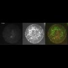
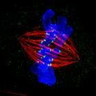
Shown is a mitotic PtK2 cell at metaphase immunostained for microtubules (red) and kinetochores (green) with DNA stained blue. The image was obtained using structured illumination microscopy (SIM, Del...
CIL:34598
NCBI Organism Classification
Homo sapiens
Biological Process
mitotic metaphase
Cellular Component
chromosome
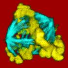
Surface representation of tripolar mitosis in a liver cancer cell line. Normally, dividing cells form a single metaphase plate of paired chromosomes which are pulled apart by opposing microtubule spi...
CIL:11988
NCBI Organism Classification
Drosophila melanogaster
Biological Process
mitotic anaphase
Cellular Component
kinesin-6
A comparison of kinesin-6 anaphase dynamics in a cell with a bipolar or monopolar spindle. (left) TIRF time lapse of kinesin-6 (Pav-GFP; green) and mCherry-tubulin (red) in a wild-type S2 cell with a ...
CIL:11990
NCBI Organism Classification
Drosophila melanogaster
Biological Process
mitotic anaphase
Cellular Component
Aurora-B
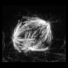
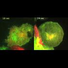
Time lapse of Aurora-B GFP and mCherry microtubules at the metaphase-to-anaphase transition. The laser illumination was set near to but not achieving complete total internal reflection TIRF to allow v...
« Previous
1
...
5
6
7
8
9
10
11
12
13
Next »
Results per page:
10
20
50
100