Alternate header for print version
Advanced search
Contributors
Help
Submit
Search
menu
Cell Process
Cell Component
Cell Type
Organism
Microbial
Alzheimer's
Data Sets
University of California, San Diego
9500 Gilman Drive
La Jolla, CA 92093-0608, USA
Voice
: (858) 534-0276
Fax
: (858) 534-7497
Email
: dorloff@ncmir.ucsd.edu
Search Results for
eukaryotic cell
(5406 results)
(Not the results you were expecting?)
(Comments)
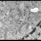
CIL:24774
NCBI Organism Classification
Rattus norvegicus
Biological Process
Golgi organization
Cellular Component
Golgi apparatus
This image is one from a set of 40nm thick serial sections through part of a Golgi ribbon from a normal rat kidney cell, generated using transmission electron microscopy following incubation at 15°C ...
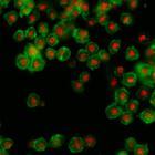
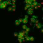
CIL:32138
NCBI Organism Classification
Drosophila melanogaster
Biological Process
cellular localization
Cellular Component
nucleus
Drosophila melanogaster Kc167 cells were stained for DNA (to label nuclei, red) and actin (a cytoskeletal protein, to show the cell body, green). Each image is a dual channel fluorescent image followe...
CIL:32143
NCBI Organism Classification
Drosophila melanogaster
Biological Process
cellular localization
Cellular Component
nucleus
Drosophila melanogaster Kc167 cells were stained for DNA (to label nuclei, red) and actin (a cytoskeletal protein, to show the cell body, green). Each image is a dual channel fluorescent image followe...
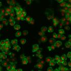
CIL:32145
NCBI Organism Classification
Drosophila melanogaster
Biological Process
cellular localization
Cellular Component
nucleus
Drosophila melanogaster Kc167 cells were stained for DNA (to label nuclei, red) and actin (a cytoskeletal protein, to show the cell body, green). Each image is a dual channel fluorescent image followe...
CIL:32131
NCBI Organism Classification
Drosophila melanogaster
Biological Process
cellular localization
Cellular Component
nucleus
Drosophila melanogaster Kc167 cells were stained for DNA (to label nuclei, red) and actin (a cytoskeletal protein, to show the cell body, green). Each image is a dual channel fluorescent image followe...
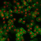
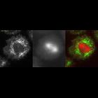
CIL:37910
NCBI Organism Classification
Homo sapiens
Biological Process
apoptotic process
Cellular Component
cortical actin cytoskeleton
Extrusion of an apoptotic cell from an HBE monolayer as shown by confocal Z-series. This set of images shows late extrusion events (early and middle events are CIL# 37908 and 37909). The bioactive lip...
CIL:25541
NCBI Organism Classification
Drosophila melanogaster
Biological Process
calcium-dependent cell-cell adhesion
Cellular Component
adherens junction
A time-lapse movie showing the dorsal pouch formation and migration of heart-anchoring cells (HANCs) in the Drosophila embryo expressing ubi-DE-cad-GFP (Shg-GFP). Shg-positive HANCs corresponding to t...
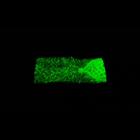
CIL:11984
NCBI Organism Classification
Drosophila melanogaster
Biological Process
mitotic anaphase
Cellular Component
myosin regulatory light chain
Time lapse of myosin GFP (left, imaged by TIRF microscopy) and mCherry tubulin (middle, imaged by epifluorescence; the spindle is close to the coverslip surface in this cell) during anaphase in a Dros...
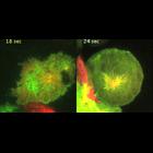
CIL:11988
NCBI Organism Classification
Drosophila melanogaster
Biological Process
mitotic anaphase
Cellular Component
kinesin-6
A comparison of kinesin-6 anaphase dynamics in a cell with a bipolar or monopolar spindle. (left) TIRF time lapse of kinesin-6 (Pav-GFP; green) and mCherry-tubulin (red) in a wild-type S2 cell with a ...
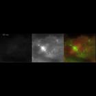
CIL:11990
NCBI Organism Classification
Drosophila melanogaster
Biological Process
mitotic anaphase
Cellular Component
Aurora-B
Time lapse of Aurora-B GFP and mCherry microtubules at the metaphase-to-anaphase transition. The laser illumination was set near to but not achieving complete total internal reflection TIRF to allow v...
« Previous
1
...
530
531
532
533
534
535
536
537
...
541
Next »
Results per page:
10
20
50
100

")