Alternate header for print version
Advanced search
Contributors
Help
Submit
Search
menu
Cell Process
Cell Component
Cell Type
Organism
Microbial
Alzheimer's
Data Sets
University of California, San Diego
9500 Gilman Drive
La Jolla, CA 92093-0608, USA
Voice
: (858) 534-0276
Fax
: (858) 534-7497
Email
: dorloff@ncmir.ucsd.edu
Search Results for
epithelial cell
(2369 results)
(Not the results you were expecting?)
(Comments)
CIL:46753
NCBI Organism Classification
Homo sapiens
Biological Process
none specified
Cellular Component
peroxisome
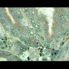
Elongated peroxisomes in duodenal epithelial cells. Varying between persons, rounded or elongated peroxisomes are observed. They also vary between epithelial cells on the villus and in the crypt. See:...
CIL:46752
NCBI Organism Classification
Mus musculus
Biological Process
peroxisome fission
Cellular Component
peroxisome
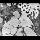
Peroxisomes in duodenal epithelial cells proliferate under treatment of oral phytol (a component of chlorophyll) over the course of 3 days. Phytol is oxidized to phytanic acid and pristanic acid, whi...
CIL:42155
NCBI Organism Classification
Mus musculus
Biological Process
dissemination
Cellular Component
plasma membrane
Representative time-lapse movie of a normal mouse mammary organoid in a 3D collagen I matrix. The cells are expressing td tomato as a membrane marker and eEGFP-actin using a keratin-14 promoter. Ima...
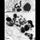
CIL:7724
NCBI Organism Classification
Rattus
Biological Process
mitosis
Cellular Component
centriole
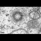
An electron micrograph showing that a centriole is constructed from a ring of nine triplet microtubules. Most animal cells contain two to four centrioles. In longitudinal view, a centriole looks like ...
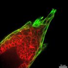
CIL:35563
NCBI Organism Classification
Rattus rattus
Biological Process
cell proliferation
Cellular Component
keratin filament
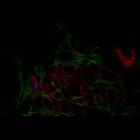
Liver of a rat exposed to chemical carcinogens. Serial sections of the liver imaged by laser scanning confocal microscopy. F-actin (red) is in bile canaliculi at junctions of the hepatocytes, which ...
CIL:48201
NCBI Organism Classification
Mus musculus
Biological Process
none specified
Cellular Component
mitochondria
Light micrograph, alcian blue PAS staining of gastric mucosa from N 18 month old gastrick H,K,ATPase null mouse, 100 x
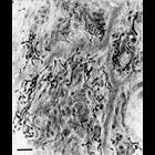
CIL:37167
NCBI Organism Classification
Rattus
Biological Process
mucus secretion
Cellular Component
none specified
Scanning electron micrograph of rat colon mucosa goblet cells. Image made available by James D. Jamieson and the Department of Cell Biology, Yale University School of Medicine.
CIL:40627
NCBI Organism Classification
Rana catesbeiana
Biological Process
detection of mechanical stimulus involved in sensory perception of sound
Cellular Component
cell surface
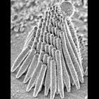
Single frog sacculus hair bundle imaged with field-emission scanning electron microscope.
CIL:47808
NCBI Organism Classification
Homo sapiens
Biological Process
none specified
Cellular Component
peroxisome
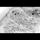
Human duodenal epithelium. Peroxisomes are stained with DAB at pH 10.5 by their catalase activity and postosmicated. 0.5 µm plastic section were visualized using electron microscopy at low magnificat...
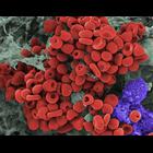
CIL:18042
NCBI Organism Classification
Mesocricetus auratus
Biological Process
erythrocyte aggregation
Cellular Component
cell
Scanning electron micrograph of a hamster oocyte cumulus complex. Cumulus cells (purple) and matrix (gray) are shown. Small blood clots (red) also often appear in oocyte cumulus complexes. The red blo...
« Previous
1
...
6
7
8
9
10
11
12
13
...
237
Next »
Results per page:
10
20
50
100


")