Alternate header for print version
Advanced search
Contributors
Help
Submit
Search
menu
Cell Process
Cell Component
Cell Type
Organism
Microbial
Alzheimer's
Data Sets
University of California, San Diego
9500 Gilman Drive
La Jolla, CA 92093-0608, USA
Voice
: (858) 534-0276
Fax
: (858) 534-7497
Email
: dorloff@ncmir.ucsd.edu
Search Results for
epithelial cell
(2369 results)
(Not the results you were expecting?)
(Comments)
CIL:25846
NCBI Organism Classification
Cricetulus griseus
Biological Process
focal adhesion disassembly
Cellular Component
focal adhesion
Time-lapse of a CHO.K1 cell cotransfected with very low levels of GFP-MIIA (myosin IIA, green) and paxillin (magenta). Yellow arrowheads mark paxillin-containing adhesions that disassemble concomitant...
CIL:11182
NCBI Organism Classification
Bufo
Biological Process
cell junction organization
Cellular Component
tight junction
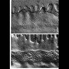
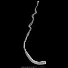
Freeze fracture replicas of zonula occludens junctions from the large intestine of a tadpole (upper) and intestine of a post-metamorphic toad (lower). The occluding junction appears as a meshwork of...
CIL:150
NCBI Organism Classification
Potorous tridactylus
Biological Process
none specified
Cellular Component
cell
Differential interference microscopy (DIC) image of PtK1 cell (marsupial kidney epithelial cell expressing dominant-negative Rac1(T17N). Live cells were used to study microtubule dynamics using X-rhod...
CIL:25357
NCBI Organism Classification
Hydra viridis
Biological Process
none specified
Cellular Component
none specified
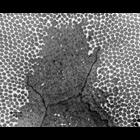
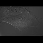
Scanning electron micrograph showing tentacles on the head of a Hydra viridis. Epithelial cells located at the base of the tentacle are in a flat, contracted state. Cell distal to the head of the H. v...
CIL:34859
NCBI Organism Classification
Homo sapiens
Biological Process
leukocyte chemotaxis involved in inflammatory response
Cellular Component
nucleus
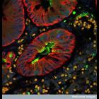
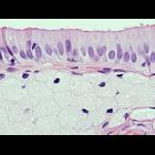
Gallbladder wall of a human patient with gallstones. The top layer is composed of highly polarized epithelial cells with microvilli partially visible at the apex and dark nuclei at the base. Below ...
CIL:35587
NCBI Organism Classification
Homo sapiens
Biological Process
mitosis
Cellular Component
spindle
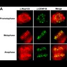
Distribution of Nup133 (a component of the Nup107-160 nucleoporin complex, red) and CENP-B (kinetochores, green) in dividing Hela cells. See Fig 2A in Orjalo et al. 2006 Mol Biol Cell 17:3806-3818.
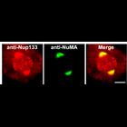
CIL:35590
NCBI Organism Classification
Homo sapiens
Biological Process
mitosis
Cellular Component
spindle
Nup133 (a component of the Nup107-160 nucleoporin complex, red) localizes to spindle poles and kinetochores while NuMA localizes to spindle poles (green) in prometaphse Hela cells. See Fig 2D in Orjal...
CIL:36148
NCBI Organism Classification
Drosophila melanogaster
Biological Process
microtubule cytoskeleton organization
Cellular Component
microtubule
The first image in this multi-image tiff file is a stimulated emission depletion image (STED) of microtubules in a Drosophila S2 cell. The second image is the corresponding diffraction limited image ...
CIL:37163
NCBI Organism Classification
Mus musculus
Biological Process
intestinal absorption
Cellular Component
microvillus
Transmission electron micrograph of a microvilli at the apex of a mouse intestine. Image made available by James D. Jamieson and the Department of Cell Biology, Yale University School of Medicine.
CIL:39016
NCBI Organism Classification
Homo sapiens
Biological Process
viral reproduction
Cellular Component
intermediate filament
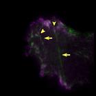
Fluorescent micrograph showing a section of human appendix infected with the measles virus. The measles virus is shown in green, cytokeratin is shown in red, which marks the epithelium, and Dapi stain...
« Previous
1
...
10
11
12
13
14
15
16
17
...
237
Next »
Results per page:
10
20
50
100


")