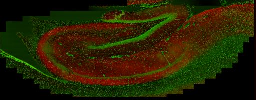
Large scale brain mosaic through the rat hippocampus immunolabeled for PMCA1a (red) and counterstained with Hoescht 33342 (green) to reveal cell nuclei. Note: hippocampus is flipped dorsal-ventral.
Localization of plasma membrane Ca2+ ATPases (PMCAs) in the rat brain
Description
Regulation of cytoplasmic calcium is crucial both for proper neuronal function and cell survival. The concentration of Ca2+ in cytoplasm of a neuron at rest is >10,000 times lower than in the extracellular space, pointing to the importance of the transporters that extrude intracellular Ca2+. The family of plasma membrane calcium-dependent ATPases (PMCAs) represent a major component of the Ca2+ regulatory system. However, little information is available on the regional and cellular distribution of these calcium pumps. We used immunohistochemistry to investigate the distribution of each of the four PMCA isoforms in the rat brain.
Funding agency
National Institutes of Health (NS51769 EES)
Leader(s)
Alain Burette
Collaborator(s)
Emanuel E. Strehler
Richard J. Weinberg
Katherine A. Kenyon
Start date
unspecified
End date
unspecified
Experiment
Experiment ID
7259
Title
PMCA1a in the adult rat brain
Purpose
To investigate the spatial distribution of the "a" variant of PMCA1 (PMCA1a) in the rat brain
Experimenter(s)
Alain Burette
Microscopy product
Microscopy product ID
7407
Instrument
RTS2000
Microscopy type
MULTIPHOTON
Product type
MOSAIC
Image basename
PMCA1A-2K-HIPPO
Spatial Axis
Image Size
Pixel Size
X
512px
0.24 um/pixels
Y
512px
0.24 um/pixels
Y
11px
Subject
Species
Rat
Scientific name
Rattus norvegicus
Strain
Sprague Dawley
Age class
adult
Tissue section
Anatomical location
Brain
Microtome
vibratome
Specimen description
Organ
brain
System
central nervous system
Imaging parameters
Type
Light microscopy product
Immersion medium
oil
Mounting medium
gelvatol
Lens
Nikon 60X oil
Lens magnification
X
Numerical aperture
1.40
Notes
ebushong
Specimen preparation
Protocol used
Brain section was immunolabeled for PMCA1a using CR1a antibody from Emanuel Strehler (1:2000). Secondary antibody was Alexa Fluor 568. Some sections were then counterstained with DAPI (Invitrogen) or Hoescht 33342 (Invitrogen) to visualized all cell nuclei and/or with NeuroTrace 640/660 (Invitrogen) to selectively visualized somata of neuronal cells. The sections were coverslipped in Gelvatol.
Imaging product type
Type
Mosaic
Description
PMCA1a labeling of hippocampus in adult rat
X position
46 tiles
Y position
19 tiles
Citation Information
Alain Burette, Emanuel E. Strehler, Richard J. Weinberg, Katherine A. Kenyon CCDB:7407, Rattus norvegicus. CIL. Dataset. https://doi.org/doi:10.7295/W9CCDB7407