Maximum intensity projection through a 3D volume of mouse brain assembled from mosaics obtained through 16 optical sections stained for fluoromyelin(green), DiI(red), and TO-PRO3(blue) which stain for myelin, blood vessels, and cells, respectively. This data is available through the Whole Brain Catalog (http://wholebraincatalog.org).
Full resolution image description
3D volume assembled from 16 optical planes from a wide field brain mosaic of coronal mouse brain tissue at 50um stained with fluoromyelin(green), DiI(red), and TO-PRO3(blue) which stain for myelin, blood vessels, and cells
Downsampled image description
downsampled coronal mouse brain tissue at 50um stained with fluoromyelin(green), DiI(red), and TO-PRO3(blue) which stain for myelin, blood vessels, and cells
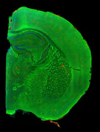
Large scale brain mosaic of a coronal section of mouse brain stained for fluoromyelin(green), DiI(red), and TO-PRO3(blue) which stain for myelin, blood vessels, and cells. Montages were assembled from each of 16 optical sections through a coronal section of mouse brain.
Full resolution image description
Coronal mouse brain tissue stained with fluoromyelin(green), DiI(red), and TO-PRO3(blue) which stain for myelin, blood vessels, and cells. Montages were assembled from each of 16 optical sections through a coronal section of mouse brain.
The Whole Brain Catalog™ is a ground-breaking, open-source, 3-D virtual environment developed by a team of researchers from UC San Diego under the Whole Brain Project™. The Catalog aims to connect members of the international neuroscience community to facilitate solutions for today's intractable challenges in brain research through cooperation and crowd sourcing. CCDB provides the backend services for very large scale image data sets on mouse brain derived from high resolution light and electron microscopy
Funding agency
Ted Waitt Family Foundation
Leader(s)
Mark Ellisman
Stephen Larson
Collaborator(s)
Sarah Maynard
Eric Bushong
Maryann Martone
Start date
08-05-2009
End date
unspecified
Experiment
Experiment ID
7281
Experiment date
10-01-2009
Title
Reconstruction of light level brain mosaics
Purpose
To reconstruct a three dimensional volume of brain slices using three different stains as reference points for reconstruction
Experimenter(s)
Eric Bushong
Monica Berlanga
Microscopy product
Microscopy product ID
7290
Instrument
Olympus Fluoview 1000
Microscopy type
LASER SCANNING CONFOCAL
Product type
MOSAIC
Image basename
3 stain_091009_T3
Subject
Species
Mouse
Scientific name
Mus Musculus
Strain
Balb C mouse
Group by
Light Microscopy
Treatment
Phosphate buffer wash and then stained with fluoromyelin, hoescht, and diI
Perfuse animal with Ringer's solution for 2 minutes followed by DiI for 10 minutes, and then 2% paraformaldehyde for 10 minutes. Remove tissue and fix overnight. Then the tissue is further dissected.3 five minute washes with cold PBS 1XStain with Fluoromyelin 1:100 overnight in cold room3 five minute washes with cold PBS 1XStain with Neurotrace 1:100 and TO-PRO3 1:3000 for 45 minutes at room tempWash once with PBS 1XStain with Neurotrace 1:100 for 45 minutes at room temperature3 five minute washes with PBS 1XMount using Gelvatol
Imaging product type
Type
Optical section
Description
Mosaics were taken at each of 16 optical sections
Z resolution
5 um
Citation Information
Mark Ellisman, Stephen Larson, Sarah Maynard, Eric Bushong, Maryann Martone (2009) CCDB:7290, Mus Musculus. CIL. Dataset. https://doi.org/doi:10.7295/W9CCDB7290