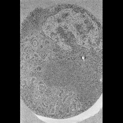
Electron micrograph of a cultured Drosophila DL1 cell infected with flock house virus, prepared by high pressure freezing followed by freeze substitution. This cell was prepared as part of an experiment to investigate different protocols for high pressure freezing.
Full resolution image description
Full sized tiff image (HPF_rec.tif) of the insect cells processed using high pressure freezing. Image corresponds to Fig. 1C in the publication.
This project is designed to achieve ultimate ultrastructure of animal tissues.
Funding agency
NIH
Leader(s)
Mark Ellisman
Gina Sosinsky
Ying Jones
Start date
01-01-2004
End date
unspecified
Experiment
Experiment ID
3469
Title
Insect
Purpose
Testing new high pressure freezing techniques on cultured cells
Experimenter(s)
Gina Sosinsky
Microscopy product
Microscopy product ID
3938
Instrument
JEOL4000EX IVEM
Microscopy type
IVEM
Product type
SURVEY
Image basename
HPF
Spatial Axis
Image Size
Pixel Size
X
5378px
Y
8013px
Subject
Species
fruitfly
Scientific name
Drosophila melanogaster
Strain
melanogaster
Group by
viral transfection
Treatment
infection with Flock House Virus
Age class
adult
Tissue section
Thickness
80 nm
Specimen description
Tissue
embryonic derived cells
Cell type
Drosophila DL1 cell
Imaging parameters
Type
Electron microscopy product
Recording medium
No recording medium provided
Magification
30000
Accelerating voltage
80 keV
Specimen preparation
Protocol used
Cell pellets were directly placed into brass planchettes that then were loaded in to the HPM 010 high pressure freezer and fast frozen. Freeze substitution: After freezing, samples (2) and (3) were placed into a Leica EM AFS Freeze substitution (FS) machine (Leica Microsystems, Bannockburn, IL) and incubated at -90 deg C for 24 hours in 0.1 percent tannic acid in acetone. Samples were washed three times with cold acetone (cooled to -90 degrees C) over 5 minutes, and placed in 1 percent OsO4 and 0.1% UA in cold acetone for 72 hours and held at -90 degrees C. After slowly warming to room temperature at 5 degrees C per hour, the specimens were rinsed in pure acetone three times (10 min. at room temperature). Infiltration and embedding in Durcupan resin was subsequently performed at room temperature.
Citation Information
Mark Ellisman, Gina Sosinsky, Ying Jones (2004) CCDB:3938, Drosophila melanogaster, Drosophila DL1 cell. CIL. Dataset. https://doi.org/doi:10.7295/W9CCDB3938