Alternate header for print version


Reconstruction
Display image description
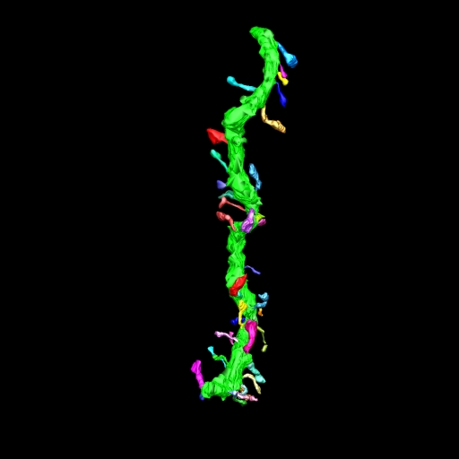
Maximum intensity projection of a tomographic reconstruction of a selectively stained spiny dendrite from a striatal medium spiny neuron from the neostriatum of a wildtype mouse.
Full resolution image description
a .tar file containing both the mrc .rec and the Analyze version of the reconstructed volume.
Volume_dimension
897, 1300, 200
Animation description
Rotation loop through a maximum intensity projection of a selectively stained spiny dendrite from a striatal medium spiny neuron from the neostriatum of a wildtype mouse. Tilt series was obtained at 2 degree increments through +/- 66 degrees of tilt.

Image 2D
Display image description
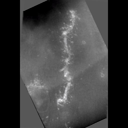
A 512 by 512 image of a zero degree tilt image of a 4 um thick section through a selectively stained spiny dendrite from a striatal medium spiny neuron in the neostriatum of a wildtype mouse.
Full resolution image description
Tar file containing IMOD files (wt_g21A2_bin3.com/.log/.st/.preali/.fid/.rawtlt) used for the alignment and the original tiff images (in the TIFF folder in the format wt_g21A2000.tif)
Animation description
Rotation loop through a maximum intensity projection of a selectively stained spiny dendrite from a striatal medium spiny neuron from the neostriatum of a wildtype mouse. Tilt series was obtained at 2 degree increments through +/-66 degrees of tilt.

Segmentation
Display image description
Spiny dendrite shaft and dendritic spines
Segmentation file description
Tar file containing the IMOD files for the segmentation (wt_g21A2_bin3.mod)

License
Attribution Only: This image is licensed under a Creative Commons Attribution License. View License Deed | View Legal Code

CCDB:3587*  Cite 
Project: P1207
Project name
Correlative microscopic characterization of dendritic spines in a transgenic mouse model of hyperdopaminergia: The dopamine transporter knockout mouse
Description
Multiscale characterization of DAT KO transgenic mouse
Funding agency
NIH
Leader(s)
Diana Price
Collaborator(s)
Aki Laakso
Michele Cyr
Maryann Martone
Naoko Yamada
Andrea Thor
Monica Berlanga
Start date
01-01-2003
End date
unspecified
 
Experiment
Experiment ID
19
Experiment date
04-22-2003
Title
P1207 Experiment 5
Purpose
EMT reconstructions of medium spiny neuron dendrites
Experimenter(s)
Diana Price
Masako Terada
Andrea Thor
Microscopy product
Microscopy product ID
3587
Instrument
Hitachi UHVEM 3MeV
Microscopy type
UHVEM
Product type
SINGLE TILT
Image basename
wt_g21A2
Subject
Species
mouse
Scientific name
Mus Musculus
Strain
C57BL/129SvJ
Group by
genetic manipulation
Treatment
NA
Age
7 months
Age class
adult
Tissue section
Anatomical location
ventral medial striatum
Microtome
ultramicrotome
Thickness
4 µm
Specimen description
Organ
brain
System
central nervous
Structure
spiny dendrite
Cell type
medium spiny neuron
Atlas coordinate
0, 0, 0,
Imaging parameters
Type
Electron microscopy product
Recording medium
film
Magification
3000
Accelerating voltage
3 MeV
Imaging product type
Type
Single tilt
Description
singlet_desc
Min range
-64 degrees
Max range
64 degrees
Tilt increment
2 degrees