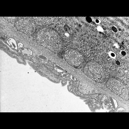
A high resolution tangential view of the surface of a non-dividing cell shows several ribbons of microtubules between the alveolar sac and the epiplasm. The thick fibrous layer associated with the epiplasm is evident, and the layer of mitochondria under the epiplasm is wrapped with rough ER. A peroxisome lies nearby and mucocysts and toxicysts are evident. TEM taken on 2/18/69 by R. Allen with Philips 300 operating at 60kV. Neg. 20,500X. The raw film was scanned with a Nikon Coolscan 9000ED. This image is suitable for quantitative analysis. Standard glutaraldehyde fixation followed by osmium tetroxide, dehydrated in alcohol and embedded in an epoxy resin. Microtome sections prepared at approximately 75nm thickness. Additional information available at (http://www5.pbrc.hawaii.edu/allen/).
| Spatial Axis | Image Size | Pixel Size |
|---|---|---|
| X | 4696px | 1nm |
| Y | 3486px | 1nm |