Licensing

Pd
Public Domain: This image is in the public domain and thus free of any copyright restrictions. However, as is the norm in scientific publishing and as a matter of courtesy, any user should credit the content provider for any public or private use of this image whenever possible. Learn more
Comments: Powered by Pnyxe

Description

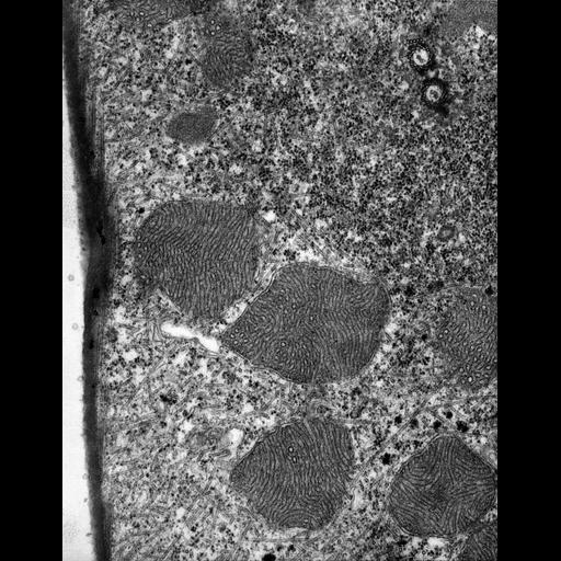
A high resolution image of the mitochondria packed with tubular cristae and the cytosol contains a jumble of microtubules. Basal bodies of the somatic kineties appear as dikinetids with a ribbon of transverse microtubules arising from the anterior basal body. The cytosol near the basal bodies is filled with ribosomes while dense rosettes of glycogen are found at the level of the mitochondria. TEM taken on 8/3/67 by R. Allen with Philips 200 operating at 60kV. Neg. 19,200X. The raw negative was scanned with an Epson Perfection V750 Pro. This image is best used for quantitative analysis. Standard glutaraldehyde fixation followed by osmium tetroxide, dehydrated in alcohol and embedded in an epoxy resin. Microtome sections prepared at approximately 75nm. Additional information is available at (http://www5.pbrc.hawaii.edu/allen/).

Biological Sources

NCBI Organism Classification
Euplotes sp.
Cell Type
cell by organism
eukaryotic cell
Eukaryotic Protist
Ciliated Protist
Cell Line
Euplotes sp. from Carolina Biological Supply
Cellular Component
mitochondrial outer membrane
mitochondrial inner membrane
symbiont-containing vacuole
glycogen granule
cytoplasmic microtubule

Biological Context

Biological Process
cortical cytoskeleton organization
mitochondrial organization
cortical microtubule organization
cytoplasm organization

Attribution

Name
Richard Allen
Link
http://www5.pbrc.hawaii.edu/allen/

Grouping

This image is part of a group.
Data Qualifiers
raw, unprocessed data
suitable for spatial measurements

Dimensions

Spatial Axis Image Size Pixel Size
X 4894px 0.78nm
Y 6302px 0.78nm
*CIL – Cell Image Library accession number. Please use this to reference an image.