Alternate header for print version


Licensing
Public Domain: This image is in the public domain and thus free of any copyright restrictions. However, as is the norm in scientific publishing and as a matter of courtesy, any user should credit the content provider for any public or private use of this image whenever possible. Learn more
tweet  
*CIL – Cell Image Library accession number. Please use this to reference an image.

CIL:9695*  Cite 
Description

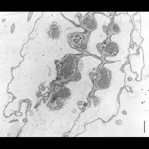
Longitudinal section of four kinetal rows of Coleps showing transverse sections of cilia and basal bodies. Parasomal sacs/clathrin coated pits penetrate through the plasma membrane into the cytoplasm adjacent to each basal body. An accumulation of early endosome-associated vesicles are near each parasomal sac. Cortical tubular mitochondria are evident. Standard glutaraldehyde fixation followed by osmium tetroxide, dehydrated in alcohol and embedded in an epoxy resin. Microtome sections prepared at approximately 75nm thickness. TEM taken on 5/23/69 by R. Allen with Philips 300 operating at 60kV. Neg. 6,370X. Bar = 1µm. A print of the negative was scanned and processed in Photoshop. This image is best used for qualitative analysis. A high resolution is available at (CIL:38827) and can be used in quantitative analysis. Additional information is available at (http://www5.pbrc.hawaii.edu/allen/).

Biological Sources
NCBI Organism Classification
Coleps hirtus
Cell Type
eukaryotic cell
Eukaryotic Protist
Ciliated Protist
Cell Line
Cambridge Culture Collection, Cambridge, England, catalog # LB1656/1
Cellular Component
plasma membrane
parasomal sacs or clathrin coated pits
alveolar membranes
cortical mitochondria
cilium
Biological Context
Biological Process
ionic transport
ciliary or flagellar motility
Molecular Function
cellular homeostasis
Attribution
Names
Richard Allen
Citation
Digital Object Identifier (DOI)
doi:10.7295/W9CIL9695
Archival Resource Key (ARK)
ark:/b7295/w9cil9695
Grouping This image is part of a group.
Imaging
Image Type
transmission electron microscopy (TEM)
illumination by electrons
Image Mode
detection of electrons
film
Parameters Imaged
electron density
Source of Contrast
stain with broad specificity
Visualization Methods
stain with broad specificity
osmium tetroxide
uranyl salt
lead salt
Processing History
recorded image
film
Print from negative scanned for Photoshop.
Data Qualifiers
processed data
suitable for spatial measurements
Dimensions
Spatial Axis Image Size Pixel Size
X 3000px ——
Y 2530px ——