Licensing
Description
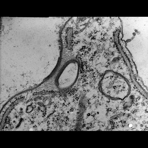
This high resolution micrograph may show important features involved in calcareous plate formation within the alveoli that underlie the plasma membrane in Coleps. Densities are seen on the inner alveolar membrane at its corners with septa. These densities are not known at this time in other ciliates and may be involved in calcareous platelet formation within the alveoli or in stabilizing the surface shape. Filamentous material fills the cytosolic space between two densities. Rough endoplasmic reticulum bears prominent ribosomes. Standard glutaraldehyde fixation followed by osmium tetroxide, dehydrated in alcohol and embedded in an epoxy resin. Microtome sections prepared at approximately 75nm thickness. TEM taken on 3/4/69 by R. Allen with Philips 300 operating at 60kV. Neg. 39,900X. The raw film was scanned with an Epson Perfection V750 Pro. This high resolution image is suitable for quantitative analysis. Additional information available at (http://www5.pbrc.hawaii.edu/allen/).
Biological Sources
- NCBI Organism Classification
- Coleps hirtus
- Cell Type
- eukaryotic cell
- Eukaryotic Protist
- Ciliated Protist
- Cell Line
- Cambridge Culture Collection, Cambridge, England, catalog # LB1656/1
- Cellular Component
- plasma membrane
- alveolar membranes
- rough endoplasmic reticulum membrane
Biological Context
- Biological Process
- membrane transport
- calcium ion transport
- Molecular Function
- calcareous plate formation
Attribution
- Name
- Richard Allen
- Link
- http://www5.pbrc.hawaii.edu/allen/
Grouping
This image is part of a group.Imaging
- Image Type
- transmission electron microscopy (TEM)
- illumination by electrons
- Imaging Mode
- detection of electrons
- film
- Parameters Imaged
- electron density
- Source of Contrast
- stain with broad specificity
- Visualization Methods
- stain with broad specificity
- osmium tetroxide
- uranyl salt
- lead salt
- Processing History
- recorded image
- film
- unprocessed raw data
- Data Qualifiers
- raw, unprocessed data
- suitable for spatial measurements
Sample Preparation
- Methods
- glutaraldehyde fixed tissue
- osmium tetroxide fixed tissue
- tissue in epoxy resin embedment
- Relation To Intact Cell
- microtome-sectioned tissue
Dimensions
| Spatial Axis | Image Size | Pixel Size |
|---|---|---|
| X | 4689px | 0.5nm |
| Y | 3624px | 0.5nm |