Licensing

Pd
Public Domain: This image is in the public domain and thus free of any copyright restrictions. However, as is the norm in scientific publishing and as a matter of courtesy, any user should credit the content provider for any public or private use of this image whenever possible. Learn more
Comments: Powered by Pnyxe

Description

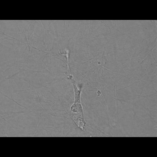
Individual Mouse Aortic Endothelial Cell (MAEC) migrating in a 3-dimensional collagen gel (1.4mg/mL). Imaged on a spinning disk confocal microscope using a 100x oil immersion objective lens on a Nikon Eclipse Ti equipped with Perfect Focus System, an electronic shutter (Smart shutter, Sutter Instruments; Novato, CA) for transmitted illumination, a linear-encoded X, Y, Z robotic stage with a piezo-driven Z-axis top plate (ASI Technologies, Montgomeryville, PA) and a Yokogawa CSU-X spinning disk confocal scan head in an electronic filterwheel. Total imaging time = 1hr, 12mins.

Biological Sources

NCBI Organism Classification
Mus musculus
(house mouse)
Cell Type
blood vessel endothelial cell
primary cell line cell
Cellular Component
cell
extracellular matrix

Biological Context

Biological Process
cell migration

Attribution

Names
Kenneth Myers
Clare M. Waterman
Other
Laboratory of Cell and Tissue Morphodynamics, NHLBI, NIH

Sample Preparation

Methods
living tissue
Relation To Intact Cell
dispersed cells in vitro
cells in 3D matrix

Dimensions

Spatial Axis Image Size Pixel Size
X 1392px 105nm
Y 1040px 105nm
Time 1 hr 12 min sec
*CIL – Cell Image Library accession number. Please use this to reference an image.