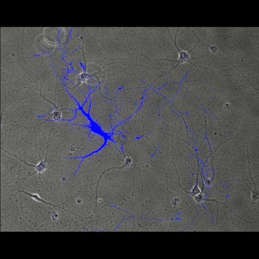
CFP-expressing neuron, 14 days in vitro. By two weeks, cultured hippocampal neurons have developed a morphology typical of mature neurons with an extensive axonal arbor, a tapered and branched dendritic arbor, and synaptic connections. Growth cones can be seen at the tips of axonal branches, and the dendritic arbor is studded with spines, specialized structures commonly associated with excitatory synapses. This image accompanies the second file in this image group which shows the fluorescent image. Embryonic rat hippocampal neurons were prepared and transfected with soluble CFP (excitation, 439; emission, 476) at plating (Dotap, Roche) as previously described (Kaech and Banker, 2006, Nat Protoc), fixed for 15 minutes (4% formaldehyde, 4% sucrose in phosphate buffered saline, pH 7.4, warmed to 37°C, permeabilized with 0.25% Triton X-100 for 7 minutes, and rinsed with phosphate buffered saline at 14 days in vitro (Withers and Banker, 1998, in Culturing Nerve Cells, MIT Press). Images were acquired with a Leica DMRXA microscope with a mercury arc lamp, a 16X PL Fluotar (0.5 NA) oil immersion lens, Princeton Instruments Micromax CCD camera and MetaMorph software, using a Leica CFP filter cube. Linked images show: A) CFP expression throughout the extent of the neuron; B) a merged image of the fluorescent cell with respect to the phase image. This image was generated with the MetaMorph overlay function, and the single plane fluorescence view is included in this image group.
| Spatial Axis | Image Size | Pixel Size |
|---|---|---|
| X | 1300px | 0.42µm |
| Y | 1030px | 0.42µm |