Licensing

Pd
Public Domain: This image is in the public domain and thus free of any copyright restrictions. However, as is the norm in scientific publishing and as a matter of courtesy, any user should credit the content provider for any public or private use of this image whenever possible. Learn more
Comments: Powered by Pnyxe

Description

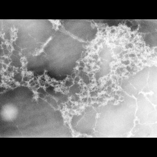
A drop of a preparation of chicken erythrocyte nuclei was exposed to low ionic strength and centrifuged onto a formvar-carbon film, fixed with paraformaldehyde and negatively stained with sodium phosphotungstate. Images were recorded at 40KX and accelerating voltage of 1MeV at the University of Wisconsin HVEM facility.This image was taken with a specimen tilt of 45 degrees. A grouped image of the same field at a 55 degree tilt provides an oblique stereo view of the tangle of chromatin fibers in the dispersed nucleus.

Biological Sources

NCBI Organism Classification
gallus gallus
Cell Type
erythrocyte
Cellular Component
nuclear chromatin

Biological Context

Biological Process
DNA packaging

Attribution

Name
Hans Ris
Pubmed
7022124

Grouping

This image is part of a group.

Sample Preparation

Methods
glutaraldehyde fixed tissue
Relation To Intact Cell
spread nucleus

Dimensions

Spatial Axis Image Size Pixel Size
X 3696px 0.5nm
Y 2791px 0.5nm
*CIL – Cell Image Library accession number. Please use this to reference an image.