Licensing

Pd
Public Domain: This image is in the public domain and thus free of any copyright restrictions. However, as is the norm in scientific publishing and as a matter of courtesy, any user should credit the content provider for any public or private use of this image whenever possible. Learn more
Comments: Powered by Pnyxe

Description

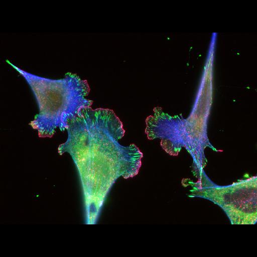
Mouse Embryonic Fibroblasts (MEFs) grown on glass coverslips coated with 10 ug/ml Fibronectin. After 24 hrs cells were fixed in 3% paraformaldehyde and stained with mouse antibody to Paxillin, rabbit antibody to cortactin, followed by anti-mouse Alexa 561 goat antibody for Cortactin (cblue), anti-rabbit Alexa 647 for Paxillin (green) and Alexa 488 phalloidin (red) to stain for F-actin. Images were collected on a Nikon Eclipse imaged with a cooled charge coupled device (CCD; Orca II; Hamamatsu Photonics) using a 60x 1.4 NA Plan Apo PH objective lens (Nikon). Individual exposure times were used for each channel: 488: 200ms, 568: 200 ms, 647: 50 ms.

Biological Sources

NCBI Organism Classification
Mus musculus
(house mouse)
Cell Type
fibroblast
primary cell line cell
Cellular Component
focal adhesion
actin cytoskeleton

Biological Context

Biological Process
cell adhesion
actin filament polymerization
Molecular Function
actin binding
cell adhesion molecule binding

Attribution

Names
Ana M. Pasapera
Clare M. Waterman
Pubmed
20308429
Other
NHLBI/ NIH

Sample Preparation

Methods
formaldehyde fixed tissue
detergent permeabilized
Relation To Intact Cell
dispersed cells in vitro

Dimensions

Spatial Axis Image Size Pixel Size
X 1392px 0.107nm
Y 1040px 0.107μm
Channel Wavelength
1 488 nm
2 568 nm
3 647 nm
*CIL – Cell Image Library accession number. Please use this to reference an image.