Licensing

Pd
Public Domain: This image is in the public domain and thus free of any copyright restrictions. However, as is the norm in scientific publishing and as a matter of courtesy, any user should credit the content provider for any public or private use of this image whenever possible. Learn more
Comments: Powered by Pnyxe

Description

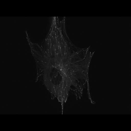
Human foreskin fibroblasts (HFF1) cells transfected with mKusabira-Orange EB3, a marker for growing microtubule ends. Note the dramatic movement of the bright fluorescent markers toward the cell periphery. Cells were imaged every 3 s by a spinning disk confocal microscoe on a Nikon Ti2000 using a 1.4NA Plan Apo Ex objective and a solid state 568 laser with a 605x52nm EM filter (Semrock). Images were recorded with a Coolsnap HQ2 (Photometrics) using 500 ms exposures.

Biological Sources

NCBI Organism Classification
Homo sapiens
(human)
Cell Type
fibroblast
primary cell line cell
Cell Line
Prepuce of penis
human foreskin fibroblast (HFF)
Cellular Component
microtubule plus end

Biological Context

Biological Process
microtubule polymerization
Molecular Function
microtubule plus-end binding

Attribution

Names
Yukako Nishimura
Clare M. Waterman
Other
NHLBI/NIH

Imaging

Image Type
recorded image
Imaging Mode
spinning disk confocal microscopy
Parameters Imaged
fluorescence emission
Source of Contrast
distribution of a specific protein
Visualization Methods
Kusabira OrangeFP
Processing History
unprocessed raw data

Sample Preparation

Methods
living tissue
Relation To Intact Cell
dispersed cells in vitro

Dimensions

Spatial Axis Image Size Pixel Size
X 1392px 0.105μm
Y 1040px 0.105μm
Time 3 sec
*CIL – Cell Image Library accession number. Please use this to reference an image.