Alternate header for print version


Licensing
Public Domain: This image is in the public domain and thus free of any copyright restrictions. However, as is the norm in scientific publishing and as a matter of courtesy, any user should credit the content provider for any public or private use of this image whenever possible. Learn more
tweet  
*CIL – Cell Image Library accession number. Please use this to reference an image.

CIL:7335*  Cite 
Description

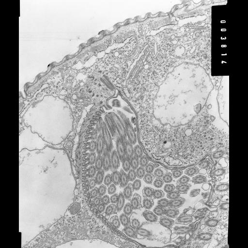
High resolution micrograph showing an overview of the peristome of Opercularia coarctata. The corrugated wall, the equivalent to the ribbed wall of Paramecium, contains tubular projections of alveoli and cross sections of two ribbons of microtubules, one of 2 microtubules and one of 4 microtubules. The ends of these ribbons, at their origins, are covered by a myoneme which, in turn, is accompanied by a layer of ER. The microtubules will extend along the wall of the peristome and some will pass beyond the cytostome to form the lamellae of the cytopharynx. Vesicles bind to the lamellae and provide membrane for the nascent food vacuole. Oral polykinetids, myoneme, endoplasmic reticulum, contractile vacuole, microtubular ribbons, pellicular pores, and Golgi complex can be seen. TEM taken on 6/10/69 by R. Allen with Philips 300 operating at 60kV. Neg. 14,800X. The raw film was scanned with an Epson Perfection V750 Pro. This image is suitable for quantitative analysis Standard glutaraldehyde fixation followed by osmium tetroxide, dehydrated in alcohol and embedded in an epoxy resin. Microtome sections prepared at approximately 75nm thickness. Additional information available at (http://www5.pbrc.hawaii.edu/allen/).

Biological Sources
NCBI Organism Classification
Opercularia [NCBITaxon:168247]
Opercularia coarctata
Cell Type
cell by organism
eukaryotic cell
Eukaryotic Protist
Ciliated Protist
Cell Line
Cambridge Culture Collection, Cambridge, England, # LB1657/1
Cellular Component
oral apparatus
contractile vacuole
microtubule basal body
Biological Context
Biological Process
ingestion
Molecular Function
intracellular organization
Attribution
Names
Richard Allen
Citation
Digital Object Identifier (DOI)
doi:10.7295/W9CIL7335
Archival Resource Key (ARK)
ark:/b7295/w9cil7335
Grouping This image is part of a group.
Imaging
Image Type
transmission electron microscopy (TEM)
illumination by electrons
Image Mode
detection of electrons
film
Parameters Imaged
electron density
Source of Contrast
stain with broad specificity
Visualization Methods
stain with broad specificity
osmium tetroxide
uranyl salt
lead salt
Processing History
recorded image
film
unprocessed raw data
Data Qualifiers
raw, unprocessed data
suitable for spatial measurements
Dimensions
Spatial Axis Image Size Pixel Size
X 3851px 1.4nm
Y 4549px 1.4nm