Alternate header for print version


Licensing
Public Domain: This image is in the public domain and thus free of any copyright restrictions. However, as is the norm in scientific publishing and as a matter of courtesy, any user should credit the content provider for any public or private use of this image whenever possible. Learn more
tweet  
*CIL – Cell Image Library accession number. Please use this to reference an image.

CIL:7329*  Cite 
Description

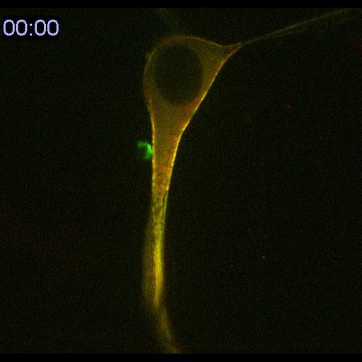
Primary endothelial cells from transgenic mouse expressing myosin heavy chain 2A fused to mCherry (red), transfected with GFP-myosin (green) regulatory light chain expression vector and demonstrate the role of myosin in cell division. Cells are embedded in 3D collagen gels. mage Name: myosin cell division timelapse Biological Source: Primary mouse (mus musculus) aortic endothelial cell. 2D images over time. 0.1274 micron/pixel, 520x490 pixels, 20 seconds interval between timepoints.Time shown is in minutes:seconds. Images collected with a spinning disk confocal on a Nikon TE1000 with a 60X 1.2NA water objective using 400 ms exposure for mCherry and 200 ms for GFP. See Fischer et al for details regarding preparation. http://www.ncbi.nlm.nih.gov/pubmed/19185493

Biological Sources
NCBI Organism Classification
Mus musculus
Cell Type
blood vessel endothelial cell
primary cell line cell
Cellular Component
myosin II complex
Biological Context
Biological Process
cell division
actomyosin contractile ring contraction
Molecular Function
myosin II binding
Attribution
Names
Robert S. Fischer
Clare M. Waterman
Pubmed
19185493
OTHER
NHLBI/NIH
Citation
Digital Object Identifier (DOI)
doi:10.7295/W9CIL7329
Archival Resource Key (ARK)
ark:/b7295/w9cil7329
Imaging
Image Type
recorded image
Image Mode
spinning disk confocal microscopy
Parameters Imaged
fluorescence emission
Source of Contrast
distribution of a specific protein
Visualization Methods
EGFP
mCherryFP
Processing History
unprocessed raw data
Sample Preparation
Methods
unfixed tissue
Methods
dispersed cells in vitro
cells in 3D matrix
Dimensions
Spatial Axis Image Size Pixel Size
X 520px 0.1274µm
Y 490px 0.1274µm
Channel Wavelength
1 488, 561nm
Time 20 seconds