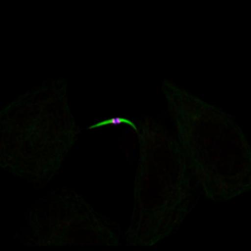
HeLa cells at last stage of cytokinesis were fixed and stained with microtubules (green), PRC1 (red), and KIF4A (blue). PRC1 and KIF4A colocalized on midbody in the center of intracellular bridge and a dark zone which cannot be accessed by antibodies was shown. Cells were fixed with MeOH on ice for 3 min, and stained with primary antibodies against PRC1 (Protein regulator of cytokinesis 1), KIF4A (Chromosome-associated kinesin), and FITC-DM1a antibody against microtubules. Secondary antibodies were Alexa 594 for PRC1, and Alexa 647 for KIF4A. A single section was collected with a spinning disk microscope on a Nikon TE-2000 with a 1.3 NA 100X objective. Images were collected with an OrCA ER CCD camera. Lasers and filters: Innova 70C Spectrum 3 watt Laser, 488 Laser/ATOF 525/50, 568 Laser/ATOF 605/52, and 647 Laser ATOF 700/75.
| Spatial Axis | Image Size | Pixel Size |
|---|---|---|
| X | 914px | 0.064µm |
| Y | 875px | 0.064µm |
| Channel | Wavelength | |
|---|---|---|
| 1 | 488nm | |
| 2 | 568nm | |
| 3 | 647nm |