Licensing

Pd
Public Domain: This image is in the public domain and thus free of any copyright restrictions. However, as is the norm in scientific publishing and as a matter of courtesy, any user should credit the content provider for any public or private use of this image whenever possible. Learn more
Comments: Powered by Pnyxe

Description

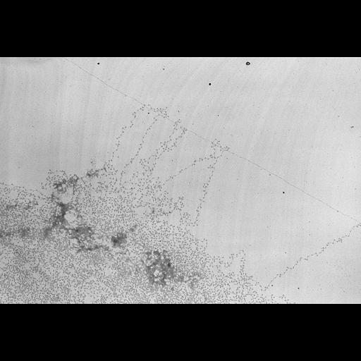
chicken erythrocyte nuclei were allowed to decondense in a low salt high pH buffer, fixed with HCOH, centrifuged onto a carbon coated EM grid, and stained with uranyl acetate. (Spreading technique of Oscar L Miller Jr). Beaded chains of nucleosomes separated by linker DNA are seen emanating from a central mass of less dispersed chromatin.

Biological Sources

NCBI Organism Classification
Gallus gallus
(red junglefowl)
Cell Type
nucleate erythrocyte
Cellular Component
chromatin

Biological Context

Biological Process
DNA packaging
Molecular Function
structural constituent of chromatin

Attribution

Name
Chris Woodcock

Imaging

Image Type
transmission electron microscopy (TEM)
Imaging Mode
illumination by electrons
Parameters Imaged
electron density
Source of Contrast
elastic scattering
Visualization Methods
visualization of contiguous regions
uranyl salt
Processing History
unprocessed raw data

Sample Preparation

Methods
formaldehyde fixed tissue
Relation To Intact Cell
isolated subcellular component

Dimensions

Spatial Axis Image Size Pixel Size
X 4096px 0.73nm
Y 2804px 0.73nm
*CIL – Cell Image Library accession number. Please use this to reference an image.