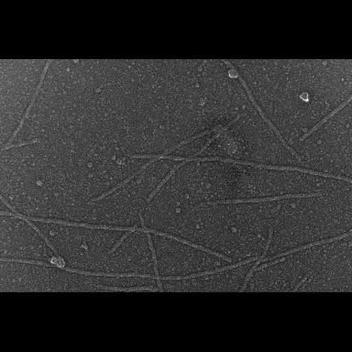
Rabbit psoas skeletal muscle fibers were blended in the presence of 5 mM MgATP to dissociate thick and thin filaments. The medium was then gently replaced with 0.3M KCl to dissociate thick filaments. Dissociated filaments were adsorbed onto mica flakes (Heuser JE, J Mol Biol. 1983 Sep 5;169(1):155-95), quick-frozen by contact with a liquid helium- cooled copper block in a Heuser-type cryopress, and freeze-etched in a Balzers 400 freeze fracture machine. The sample was then rotary-replicated with Pt-C at 11 degrees and visualized in a JEOL 100CX2 transmission electron microscope operated at 100 kev. The image was recorded on Kodak 4489 film at 100,000x magnification. Stereo pairs were taken at +5 and -5 degrees. Films were scanned with a 20 micrometer pixel spacing on a Nikon Coolscan 9000ED scanner. Stereo pair with CIL:6262.
| Spatial Axis | Image Size | Pixel Size |
|---|---|---|
| X | 4142px | 0.00027µm |
| Y | 2749px | 0.00027µm |