Alternate header for print version


Licensing
Public Domain: This image is in the public domain and thus free of any copyright restrictions. However, as is the norm in scientific publishing and as a matter of courtesy, any user should credit the content provider for any public or private use of this image whenever possible. Learn more
tweet  
*CIL – Cell Image Library accession number. Please use this to reference an image.

CIL:6230*  Cite 
Description

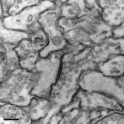
This study on the zebrafish optic tectum is a series of four images with magnifications ranging from the SEM block face overview to of the zebrafish head to high magnification (80,000X) showing optic tectum synapse. The first image is a bloc face SEM image at 200x of the epon embedded zebrafish head. The second image is another block face SEM low magnification image of the tectum. The third image is a 60,000X TEM image of a OTO fixation and copper lead en bloc staining of zebrafish optic tectum synapse. This image, the fourth image in the series, is a 80,000X TEM image of a zebrafish optic tectum synapse. Zebrafish 10 dpf Optic Tectum Synapses. Image collected on a JOEL 1230 at 80 kV using a Gatan 967 slow-scan, cooled CCD camera at 80,000X. 70 nm thick section. Microwave processing, osmium tetroxoxide/osmid procedure for enhancing membranes, and copper lead en bloc staining. Procedure for specimen preparation available at http://www.stanford.edu/~redhair/JoAnn_Buchanan.

Biological Sources
NCBI Organism Classification
Danio rerio
Cell Type
CNS neuron (sensu Vertebrata)
Cellular Component
synapse
Biological Context
Biological Process
synapse organization
Zebrafish Anatomy
optic tectum
Attribution
Names
Buchanan, JoAnn (Stanford University School of Medicine)
Link
http://www.stanford.edu/~redhair/JoAnn_Buchan...
Citation
Digital Object Identifier (DOI)
doi:10.7295/W9CIL6230
Archival Resource Key (ARK)
ark:/b7295/w9cil6230
Grouping This image is part of a group.
Imaging
Image Type
recorded image
Parameters Imaged
electron density
Source of Contrast
differences in adsorption or binding of stain
Visualization Methods
osmium tetroxide
lead salt
copper salt
Processing History
unprocessed raw data
Dimensions
Spatial Axis Image Size Pixel Size
X 1024px 1.6nm
Y 1024px 1.6nm