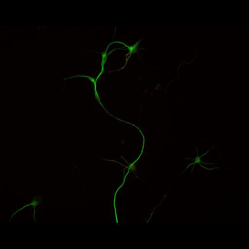
Synapse formation in cultured hippocampal neurons after 7 days in vitro. Cells were immunostained for MAP2, a microtubule associated protein localized to dendrites but not axons (green), and synapsin I, a presynaptic vesicle protein (red) to reveal the development of presynaptic contacts along the dendritic arbor.
Embryonic rat hippocampal neurons were prepared as previously described (see Kaech and Banker, 2006, Nat Protoc). Cells were prepared for fluorescent staining as previously described (Withers and Banker, 1998, in Culturing Nerve Cells, MIT Press). Briefly, cells were fixed (4% formaldehyde, 4% sucrose in phosphate buffered saline, pH 7.4), permeabilized with 0.25% Triton and immunostained MAP2 (HM2, from Sigma with d549 conjugated secondary, excitation, 555, emission, 568, Jackson Immunoresearch). Images were acquired with a Leica DMRA microscope with a 16X PL Fluotar (0.5 NA) lens, Photometrics CoolSnap ES CCD camera and MetaMorph software. This merged image was generated using the color combine function in Metamorph.
| Spatial Axis | Image Size | Pixel Size |
|---|---|---|
| X | 1300px | 0.42µm |
| Y | 1030px | 0.42µm |