Alternate header for print version


Licensing
Attribution Non-Commercial Share Alike:This image is licensed under a Creative Commons Attribution, Non-Commercial Share Alike License. View License Deed | View Legal Code
tweet  
*CIL – Cell Image Library accession number. Please use this to reference an image.

CIL:54890*  Cite 
Description

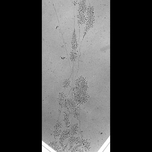
Part of a collage of images of a single nucleolus from yeast strain NOY1051 which has a fixed rDNA array size of 143 repeats. In total 71 of 120 35S ribosomal RNA genes visualized in the Miller chromatin spread from this cell are transcriptionally active. An overview of this nucleolus photographed at low magnification is published in French et al. (2008) Mol Cell Biol. 28:4576-87, Fig. 3A. https://www.ncbi.nlm.nih.gov/pmc/articles/PMC2447126/figure/f3/

Technical Details

Miller chromatin spread. For yeast technique see: Osheim YN, French SL, Sikes ML, Beyer AL. Electron microscope visualization of RNA transcription and processing in Saccharomyces cerevisiae by Miller chromatin spreading. Methods Mol Biol. 2009;464:55-69. doi: 10.1007/978-1-60327-461-6_4. PMID: 18951179.

Biological Sources
NCBI Organism Classification
Saccharomyces cerevisiae
Cell Type
Single cell
Cellular Component
nucleolus
Biological Context
Biological Process
Transcription (RNA polymerase I)
Attribution
Names
Sarah French
Link
Pubmed
OTHER
Cells grown at 30 degC in rich medium were harvested in mid-log phase.
Citation
Digital Object Identifier (DOI)
doi:10.7295/W9CIL54890
Archival Resource Key (ARK)
ark:/b7295/w9cil54890
Grouping This image is part of a group.
Imaging
Image Mode
transmission electron microscopy (TEM)
Visualization Methods
Uranyl acetate
phosphotungstic acid
Sample Preparation
Methods
Image was captured at a magnification of 6600X on electron microscope film, then enlarged an additional 3.25X when printed onto photographic paper and scanned at 600 dpi.
Dimensions
Spatial Axis Image Size Pixel Size
X 2069px 1.97nm
Y 4587px 1.97µm