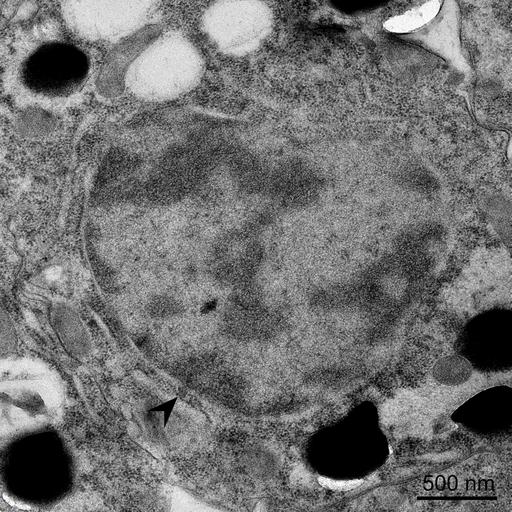
Wild type Caenorhabditis elegans (N2 background) were high pressure frozen and visualized through transmission electron microscopy (TEM). The images show the nucleus of intestine cells and Oocytes and the black arrow indicates nuclear envelope budding events.
Worms were cultured onto normal growing medium plates spread with Escherichia coli (OP50 strain) and high pressure frozen in a Wohlwend Compact 3 machine, followed by a short freeze substitution protocol in 2% Uranyl acetate and embedded in HM20 resin. Sections of 70 nm thickness were then contrast stained with 2% Uranyl acetate and Reynold's lead citrate. Pictures were aquired in a Tecnai T12 TEM with a Ceta CMOS 16M camera.