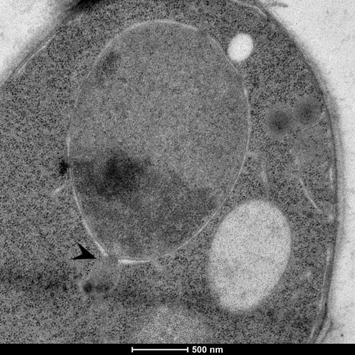
Young Saccharomyces cerevisiae cells were collected after separation of the old cells from the culture. Old cells were isolated with the use of Streptavidin magnetic beads and the remaining young population was high pressure frozen and visualized with the use of transmission electron microscopy (TEM). The images show the nucleus of the cells and the black arrow indicates nuclear envelope budding events.
The cell surface of exponentially growing Saccharomyces cerevisiae cells was labeled with biotin by incubating the cells with 0,5 mg/ml Sulfo-NHS-LC biotin (#21335, Thermo Fisher scientific) for 20 minutes at room temperature. Cells were grown in YPD, harvested prior saturation of culture, and washed in PBS + 0,5% glucose. Biotinylated cells were labeled with 17.5 ug/ml Streptavidin magnetic beads (#21344, Thermo Fisher scientific) for 1.5 hours followed by 3 x 15 min magnetic sorting with PBS + 0,5% glucose washes in between. Another two rounds of growth, streptavidin-labeling, and sorting was performed. The unbound daughter cells were recovered in YPD for 4 hours before high pressure freezing. Samples were high pressure frozen in a Wohlwend Compact 3 machine, followed by a short freeze substitution protocol in 2% Uranyl acetate and embedded in HM20 resin. Sections of 70 nm thickness were then contrast stained with 2% Uranyl acetate and Reynold's lead citrate. Pictures were aquired in a Tecnai T12 TEM with a Ceta CMOS 16M camera.