As biological imaging datasets increase in size, deep neural networks are considered vital tools for efficient image segmentation. While a number of different network architectures have been developed for segmenting even the most challenging biological images, community access is still limited by the difficulty of setting up complex computational environments and processing pipelines, and the availability of compute resources. Here, we address these bottlenecks, providing a ready-to-use image segmentation solution for any lab, with a pre-configured, publicly available, cloud-based deep convolutional neural network on Amazon Web Services (AWS). We provide simple instructions for training and applying CDeep3M for segmentation of large and complex 2D and 3D microscopy datasets of diverse biomedical imaging modalities.
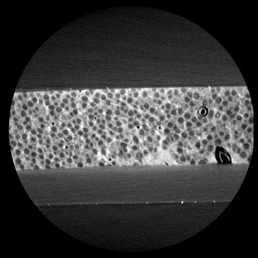
Zeiss Xradia 510 Versa (Zeiss X-Ray Microscopy) operated at 40 kV (76 µA current) with ×40 magnification . Tilt series of 3201 projections using XMReconstructor (Xradia). Mouse brain, hippocampal section, from the center of the suprapyramidal blade of the dentate gyrus (DG) to the molecular layer
| Spatial Axis | Image Size | Pixel Size |
|---|---|---|
| X | 940px | 391µm |
| Y | 974px | 405µm |
| Z | 948px | 395µm |