Alternate header for print version


Licensing
Attribution Non-Commercial Share Alike:This image is licensed under a Creative Commons Attribution, Non-Commercial Share Alike License. View License Deed | View Legal Code
tweet  
*CIL – Cell Image Library accession number. Please use this to reference an image.

CIL:50636*  Cite 
Description

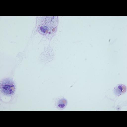
Two monocytes with cytoplasmic erythrocytes were detected (upper part and lower right corner of the photo). The two other cells show erythrocytes at contact (one of which also a rectangular crystal). To note would be that for one of them, the erythrocytes are situtated between 2 monocytes, for the other, the erythrocytes are covered by cytoplasmic-appearing material (possibly from the respective monocyte or from another).

Technical Details

Cerebrospinal fluid, cytocentrifugation, May-Grunwald-Giemsa stain.

Biological Sources
NCBI Organism Classification
Homo sapiens
Cell Type
monocyte
erythrocyte
Cellular Component
Cytoplasm lesion
Biological Context
Biological Process
Erytrophagocytosis
Attribution
Names
Adriana Handra-Luca,
APHP University Paris Nord
Citation
Digital Object Identifier (DOI)
doi:10.7295/W9CIL50636
Archival Resource Key (ARK)
ark:/b7295/w9cil50636
Imaging
Image Mode
microscopy
Visualization Methods
x100